Case Blog - (2015) Volume 3, Issue 2
Divya Arora1* and Adi Singh2 |
Corresponding Author: Divya Arora, Baylor Scott and White Hospital, Department of Radiation Oncology 2401, South 31st Street, Temple, TX 76508, USA,Tel: 254-724-7091,E-mail: arorad86@gmail.com |
Related article at Pubmed, Scholar Google |
A 9 year old patient who initially presented with headaches was found to have a high risk medulloblastoma of the cerebellar vermis. He presented status post craniotomy and subtotal resection. The patient developed posterior fossa syndrome with ataxia, dysmetria, and cerebellar mutism. He underwent craniospinal radiation therapy with concurrent vincristine. During radiation, he presented with worsening headaches and was found to have drop metastasis in the spine with increased residual tumor in the posterior fossa. This was confirmed via MRI of the brain, thoracic spine, and lumbar spine. He continued radiation to a total dose of 36 Gray to the whole brain and spine with a boost to the posterior fossa. He was started on maintenance therapy per ACNS0332 protocol (without isoretinoin). After three months of radiation he showed response to therapy on repeat images. Two months later, he presented with worsening headaches, emesis, watery diarrhea and colicky abdominal pain. Imaging at this time revealed leptomeningeal spread of his medulloblastoma with involvement of the cervical, thoracic and lumbar spine. Brain MRI showed leptomeningeal disease with intraventricular carcinomatosis with extension into the left caudate and periventricular white matter. After progression was confirmed, the patient’s family elected for home hospice. One week later, the patient was deceased. MRI
A 9 year old patient who initially presented with headaches was found to have a high risk medulloblastoma of the cerebellar vermis. He presented status post craniotomy and subtotal resection. The patient developed posterior fossa syndrome with ataxia, dysmetria, and cerebellar mutism. He underwent craniospinal radiation therapy with concurrent vincristine. During radiation, he presented with worsening headaches and was found to have drop metastasis in the spine with increased residual tumor in the posterior fossa. This was confirmed via MRI of the brain, thoracic spine, and lumbar spine. He continued radiation to a total dose of 36 Gray to the whole brain and spine with a boost to the posterior fossa. He was started on maintenance therapy per ACNS0332 protocol (without isoretinoin). After three months of radiation he showed response to therapy on repeat images. Two months later, he presented with worsening headaches, emesis, watery diarrhea and colicky abdominal pain. Imaging at this time revealed leptomeningeal spread of his medulloblastoma with involvement of the cervical, thoracic and lumbar spine. Brain MRI showed leptomeningeal disease with intraventricular carcinomatosis with extension into the left caudate and periventricular white matter. After progression was confirmed, the patient’s family elected for home hospice. One week later, the patient was deceased. |
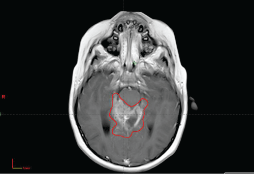
MRI Brain on Initial Presesentation with Gross Tumor Volume (GTV) Contoured for Radiation Planning. |
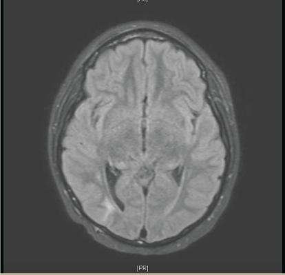
MRI Brain showing interval response two months after chemotherapy and craniospinal radiation (36 Gy). |
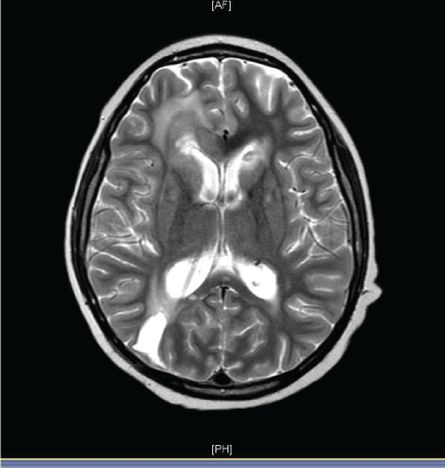
MRI Brain at time of progression five months after radiation. |
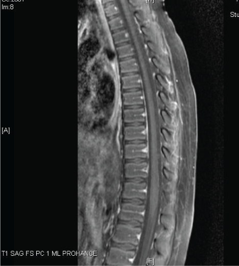
MRI Thoracic spine at initial presentation showing no evidence of metastasis. |
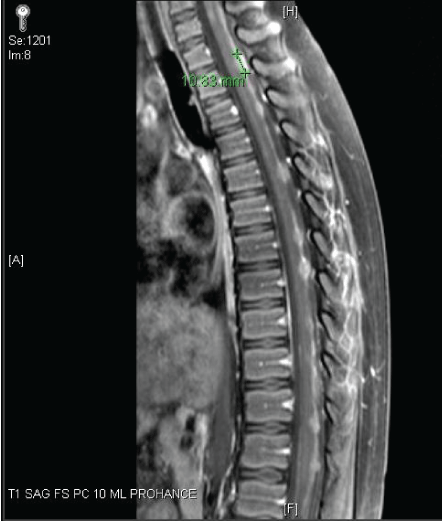
MRI Thoracic spine one month later with drop metastasis while on craniospinal radiation. |