Image - (2016) Volume 4, Issue 2
A Rare Case of Global Developmental Delay with Ataxia Joubert Syndrome
ANagesh Dasarwar* and Sravya Datla
Consultant Pediatrician and Neonatologist, GMR Varalaxmi Care Hospital, Andhra Pradesh, India
*Corresponding Author:
Nagesh Dasarwar
Consultant Paediatrician and Neonatologist
GMR Varalaxmi Care Hospital, Rajam, Dist Srikakulam
Andhra Pradesh
India
Tel: 080042 51834
E-mail: vivek03sharma@rediffmail.com
Received date: April 11, 2016; Accepted date: April 13, 2016; Published date: April 16, 2016
Citation: Dasarwar N, Datla S. A Rare Case of Global Developmental Delay with Ataxia: Joubert Syndrome. Ann Clin Lab Res. 2015, 4:2.
Abstract
Joubert syndrome is a rare autosomal recessive disorder characterised by developmental delay, ataxia, episodic hyperapneoa, opsoclonus associated with brainstem and cerebellar abnormalities. We describe clinical and neuroimaging features of a child with classical joubert syndrome. A 22 months old male child born of non-consanguineous marriage, presented to us with global developmental delay, hypotonia, truncal ataxia and abnormal eye movements. Birth history was uneventful. MRI brain revealed classical features of joubert syndrome. The MRI features are thickened superior cerebellar peduncles, hypoplasia of the vermis, giving it an appearance of molar tooth sign. The molar tooth sign is almost pathognomonic of joubert syndrome. The prognosis of these patients is poor with a five year survival rate of less than 50%. As the recurrence rate is 25% prenatal diagnosis should be done using serial ultrasounds and foetal MRI in second trimester. These children are sensitive to respiratory depressant effects of opiates and nitrous oxide, hence should be avoided
Joubert syndrome is a rare autosomal recessive disorder characterised by developmental delay, ataxia, episodic hyperapneoa, opsoclonus associated with brainstem and cerebellar abnormalities.
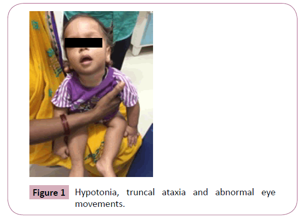
We describe clinical and neuroimaging features of a child with classical joubert syndrome. A 22 months old male child born of non-consanguineous marriage, presented to us with global developmental delay, hypotonia, truncal ataxia and abnormal eye movements (Figure 1). Birth history was uneventful.

Figure 1: Hypotonia, truncal ataxia and abnormal eye movements.
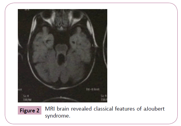
MRI brain revealed classical features of Joubert syndrome. The MRI features are thickened superior cerebellar peduncles, hypoplasia of the vermis, giving it an appearance of molar tooth sign (Figure 2).The molar tooth sign is almost pathognomonic of Joubert syndrome.

Figure 2: MRI brain revealed classical features of aJoubert syndrome.
The prognosis of these patients is poor with a five year survival rate of less than 50%. As the recurrence rate is 25% prenatal diagnosis should be done using serial ultrasounds and foetal MRI in second trimester. These children are sensitive to respiratory depressant effects of opiates and nitrous oxide, hence should be avoided.
9034


