Case Blog - (2017) Volume 5, Issue 1
An Atypical Squamous Cell Carcinoma of the Cervix
Sarah El-Abbassi*, Asmae Touil, Tayeb Kebdani and Noureddine Benjaafar
Department of Radiation Oncology, National Intitute of Oncology, Mohammed V University, Rabat, Morocco
*Corresponding Author:
Sarah El-Abbassi
Resident, Department of Radiation Oncology
National Institute of Oncology
Mohamed V University, Rabat, Morocco
Tel: +212661678389
E-mail: drsarahelabbassi@gmail.com
Received date: 17 February 2017; Accepted date: 27 February 2017; Published date: 03 March 2017
Citation: El-Abbassi S, Touil A, Kebdani T, et al. An Atypical Squamous Cell Carcinoma of the Cervix. Arch Can Res. 2017, 5:1. doi:10.21767/2254-6081.1000135
Abstract
A 74-year-old female presented in our department with 4 months history of post-menopausal bleeding and pelvic pain, which was increasing in intensity over 2 months. However, she denied any weight loss, fever or altered urinary or bowel habits. Her previous medical history revealed hypertension since 10 years for that she was taking antihypertensive drugs. On physical assessment she was not pale, with good performance status. Per vaginal examination showed an ulcer and budding tumor destroying the cervix and the the upper vagina.
Case Blog
A 74-year-old female presented in our department with 4 months history of post-menopausal bleeding and pelvic pain, which was increasing in intensity over 2 months. However, she denied any weight loss, fever or altered urinary or bowel habits. Her previous medical history revealed hypertension since 10 years for that she was taking antihypertensive drugs. On physical assessment she was not pale, with good performance status. Per vaginal examination showed an ulcer and budding tumor destroying the cervix and the the upper vagina.
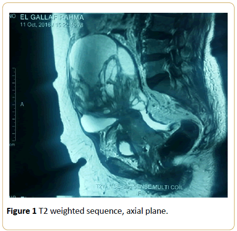
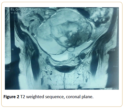
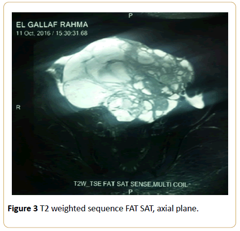
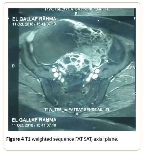
A cervical biopsy was taken. It demonstrated well and invasive squamous cell carcinoma. Magnetic Resonance Imaging (MRI) showed a big tumor measuring 10.4 cm × 21 cm × 17 cm, extending forward to the bladder, up to the uterus and the anterior abdominal wall (Figures 1-4). With evidence of parametriale invasion and bilateral pelvic lymphadenopathy: 2 left internal iliac enlarged lymph nodes. The largest was measuring about 1.5 cm short axis. She was staging according to the International Federation of Gynecology and Obstetrics staging system 2009 as FIGO IVa. After multidisciplinary board meeting, patient started chemotherapy. No chemoradiation was done because of tumor size.

Figure 1: T2 weighted sequence, axial plane.

Figure 2: T2 weighted sequence, coronal plane.

Figure 3: T2 weighted sequence FAT SAT, axial plane.

Figure 4: T1 weighted sequence FAT SAT, axial plane.
Figures 1 - 4 show the MRI, an appearance of cervical squamous cell carcinoma in different planes and sequences.
18428


