Chloë Fidanzi-Dugas, Bertrand Liagre*, Guillaume Chemin, Aurélie Perraud, Claire Carrion, Robert Granet, Vincent Sol and David Yannick Léger
Department of Biochemistry and Molecular Biology, 2 Rue du Dr Raymond Marcland, 87025 Limoges, France
Corresponding Author:
Bertrand Liagre
Department of Biochemistry and Molecular Biology
2 Rue du Dr Raymond Marcland, 87025 Limoges, France
Tel: 33 5 55 43 58 00
E-mail: bertrand.liagre@unilim.fr
Received date: 21 July 2016; Accepted date: 23 July 2016; Published date: 26 July 2016
Citation: Fidanzi-Dugas C, Liagre B, Chemin G, et al. Analysis of the In Vitro and In Vivo Effects of the Photodynamic Therapy on Prostate Cancer by using News Photosensitizers, Protoporphyrin IX- Polyamines Derivatives. Arch Can Res. 2016, 4:3.
To determine potential mechanisms by which PpIX-PA (protoporphyrin IX-polyamines; PpIX is coupled with two molecules of spermidine (PpIX-dSd) or spermine (PpIX-dSm)) inhibited prostate cancer cell viability, we studied its effects on mitochondrial membrane potential because alterations in mitochondrial structure and function have been shown to play a crucial role at early stages of apoptosis
Image Case
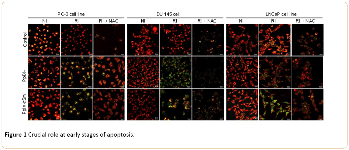
To determine potential mechanisms by which PpIX-PA (protoporphyrin IX-polyamines; PpIX is coupled with two molecules of spermidine (PpIX-dSd) or spermine (PpIX-dSm)) inhibited prostate cancer cell viability, we studied its effects on mitochondrial membrane potential because alterations in mitochondrial structure and function have been shown to play a crucial role at early stages of apoptosis (Figure 1).

Figure 1: Crucial role at early stages of apoptosis.
PpIX-PA induced apoptosis via the intrinsic pathway in prostate cancer cell lines PC-3, DU 145 and LNCaP. Cells were cultured in 10% FCS medium during 24 hrs, treated or not with PpIX-PA (IC50) for 24 hrs, treated or not with NAC (10 mM) and irradiated (RI) or not (NI) by red light (75 J/cm2;). After 24 hrs, cells were incubated with medium containing JC-1 (1 μg/ml) for 30 min at 37°C. Red fluorescence represents mitochondria with intact membrane potential whereas green fluorescence represents de-energized mitochondria. Pictures were taken with a confocal microscope (laser Zeiss LSM 510 Meta - X200). The pictures are representative of two separate experiments.
9955


