Hassan R AL-Rikabi1 , Hayder Hussein Jalood2* and Ali Juma Mohammed3
1Department of Biology, College of Pure Education, University of Thi Qar, Nasiriyah, Iraq
2Department of Pharmacy, Mazaya University College, Baghdad, Iraq
3Ministry of Education, General Directorate of Education in Thi-Qar, Iraq
*Corresponding Author:
Hayder Hussein Jalood
Department of Pharmacy, Mazaya University College
Baghdad, Iraq
Tel: 07831072028
E-mail: mtqr86@gmail.com
Received Date: March 31, 2017; Accepted Date: May 04, 2017; Published Date: May 11, 2017
Citation: AL-Rikabi HR, Jalood HH, Mohammed AJ. Association of C-469T of Interleukin 13 Gene Polymorphism with Vitiligo Development in Thi Qar Province/South of Iraq. J Biomedical Sci. 2017, 6:3. doi: 10.4172/2254-609X.100064
Copyright: © 2017 AL-Rikabi HR, et al. This is an open-access article distributed under the terms of the Creative Commons Attribution License, which permits unrestricted use, distribution, and reproduction in any medium, provided the original author and source are credited.
Keywords
Interleukin; Polymorphism; Vitiligo; PCR-RFLP
Introduction
Vitiligo is an acquired disorder characterized by the appearance of white patches resulting from the loss of melanocytes and melanin from the skin. This disorder affects 1– 4% of the world population. There are several hypothesis concerning the etiology of vitiligo have been suggested including autoimmunity, inherent defects in melanocyte biochemistry and neuronal dysfunction [1]. Among these, believes the most well accepted is the autoimmune theory, supported by findings that are proven the frequent occurrence of other autoimmune diseases in vitiligo patients and their relatives [2] also the presence of auto-antibodies and auto-reactive T-lymphocytes in vitiligo patients, both with cytotoxic effects upon melanocytes [3]. The immune system is complex, involving cell-mediated and humoral mechanisms – both of which appear to play roles in the manifestation of vitiligo. Identifying pathways involved in the immune reactions in vitiligo will help in understanding the cause of vitiligo and pave the way for developing specific immune targets to combat the disease. Co-occurrence with autoimmune diseases seems to be dependent of the clinical type of vitiligo especially with nonsegmental type [2]; however, the observation is not as frequent when considering exclusively the segmental clinical type of vitiligo [4]. The changing in cytokines levels were reported in vitiligo affected skin compared with healthy skin suggesting that the cytokine production in epidermal microenvironment may play a role in pathogenesis of vitiligo [5,6]. Several studies showed the role of peripheral blood and lesional cytokine expression in vitiligo patients. These suggested a role for epidermal cytokine imbalance in the pathogenesis of vitiligo [6,7].
Increased levels of a class of proteins called interleukins (ILs) might be linked to the active stage of vitiligo [8]. Several studies investigated the putative role of cytokines in vitiligo by studying IL-17 [9] , IL-10 [7], IL-2,IL-6 [10]. IL-6 and IL-13 secreting CD8+ T cells from vitiligo perilesional margins may induce autologous melanocyte apoptosis [11]. Increased concentrations of serum IL-10, IL-13, and IL-17A and decreased concentrations of TGF-β1 suggested altered cell-mediated immunity that may facilitate the melanocyte cytotoxicity in vitiligo [8].
Human Interleukin-13 (IL-13) is a 17-KDa glycoprotein. IL-13 is produced mainly by activated Th2 cells [12] and is contributed in the maturation and differentiation of B cells [13]. The IL-13 gene is located on chromosome 5q31 in the cluster of genes encoding IL-3, IL-4, IL-5 and IL-9. It has four exons and five introns [14]. More than thirty one single nucleotide polymorphisms have been identified in the IL-13 gene of human. Most of these polymorphisms are in the 5 regulatory region or in intron (noncoding region) of the gene. The most widely studied SNPs are promoter polymorphism T-1112C and anonsynonymous polymorphism located in exon A-2044C. Functional studies have explained the association between the IL-13 SNPs and phenotype of some diseases such as asthma [15,16] , diabetes [17].
Subjects and Methods
In this study sixty patients with vitiligo disease were randomly selected. The subjects composed of 40 males and 20 females, whose ages ranged from (1–58) years. They were from clinic of dermatology of Al-Hussein Teaching Hospital in the province of Thi Qar. Forty healthy cases were studied as matched controls. All the cases studied had no other autoimmune diseases. Blood samples were collected from all subjects into an EDTA vacutainer tubes (2.5 ml). Genomic DNA was extracted from blood leukocytes using a spin column protocol [18] by gSYNCTMDNA Mini kit.
C-469T Polymorphism
The C-469T polymorphism in the IL-13 gene was detected by a polymerase chain reaction –restriction fragment length polymorphism (PCR-RFLP) method was suggested by Graves and Coworkers [19]. The following primers were used in PCR technique, F- 5-CCT AGG CAG GCA ACA TAG TG-3 and R-5-CTG GAC CCT TCT CAA TAA GT-3. PCR was carried out in a total volume of 20 μl with 5 μl of DNA, 1 μl of each primers, 5 μl master mix, 9 μl distal water .The amplification conditions were explained in Table 1.
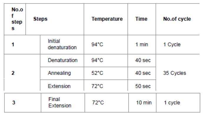
Table 1: Program of polymerase chain reaction technique.
RFLP Analysis
The products of PCR were cleaved with AccI as follows: 5 μl of the PCR product were added to 1 μl of the enzyme and then 2 μl buffer and 10 μl sterile distilled water were added, followed by incubation at 37°C for 2 hours and then electrophoresed on 2% agarose stained with ethedium bromide.Statistical Analysis
Statistical Analysis
Statistical analysis of this study was conducted, using the mean, standard deviation, chi-square test and odd ratio test with 95% confidence intervals (95% CI) by SPSS V.17.
Results
Clinical features were showed in Table 2. In this study, the mean age of patient group was 31.85 ± 15.1 years and the mean of age onset was 20.9 ± 13.7. Regarding to gender, 66.67% of patients were male while 33.33% were female. 68.33% of cases were non-smokers and 31.37% were smokers. 35% of patients have family history, meanwhile the rest have no family history.
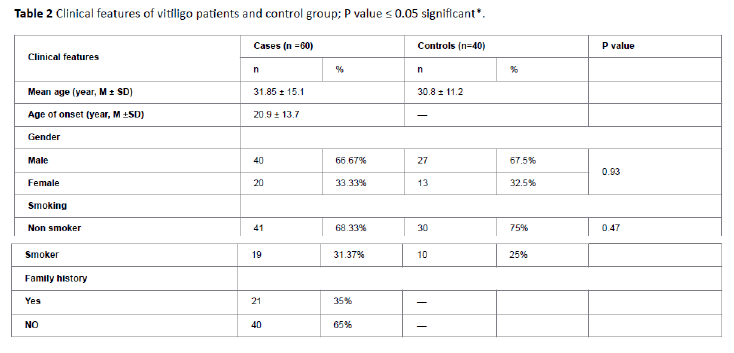
Table 2: Clinical features of vitiligo patients and control group; P value ≤ 0.05 significant*.
Amplification of flanking regions of C-469T resulted in 549 bp, 303 bp and 246 bp bands. The PCR-RFLP analysis for C-469T SNP generated two fragments in case of TT genotype and the fragments were 303 bp and 246 bp. As for CC genotype a 549 bp was seen. On the other hand , all three bands 549/303/246 bp could be showed in case CT genotype.
For C-469T SNP, the results showed no significant of CT genotype P=0.48 (OR=1.4; 95 %CI=0.53-3.75) in patients compared to control group. Also TT genotype was no significantly more frequent in patients P=0.59 (OR=0.76; 95% CI=0.28-2) (Table 3). T allele frequent did not show significant compared to C allele in patient and control groups P=0.6 (OR=0.85 ; 95 %CI=0.48 - 1.52) (Table 4).
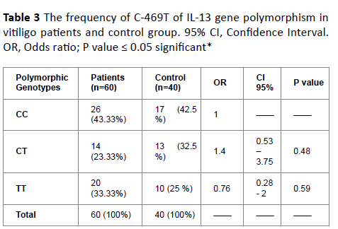
Table 3: The frequency of C-469T of IL-13 gene polymorphism in vitiligo patients and control group. 95% CI, Confidence Interval. OR, Odds ratio; P value ≤ 0.05 significant*
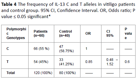
Table 4: The frequency of IL-13 C and T alleles in vitiligo patients and control group. 95% CI, Confidence Interval. OR, Odds ratio; P value ≤ 0.05 significant*
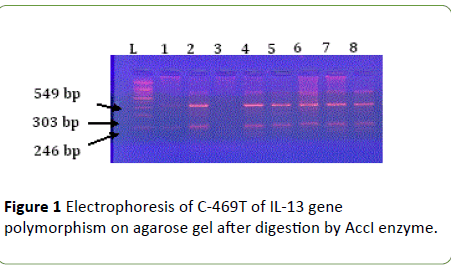
Figure 1: Electrophoresis of C-469T of IL-13 gene polymorphism on agarose gel after digestion by AccI enzyme.
Discussion
No significant differences were observed between the two studied groups as regards sex, age and smoking. The mean age was 31.85 ± 15.1 years and 30.8 ± 11.2 years of cases and control group respectively. This finding is similar to Usha and Pandey [20] who found the mean age of patient and control group was 33.23 ± 16.67 and 31.45 ± 11.02 respectively. Age of onset of disease was 20.9 ± 13.7. Most of studies demonstrated the onset of vitiligo was generally at less than 30 years in age [21,22]. Vitiligo does not show any significant association with gender (P=0.10). Although the majority of patients were male (66.67%), but we found no significant in gender between the vitiligo patients and the control group (P=0.93). Other study showed that disease was more common in female [21]. We found no significant between patient and control group according to smoking (P=0.47). This finding suggested by [20,23]. A positive family history appeared in 35 % of patients. Also [23] found 30% of cases had positive family history. Less than that [24] reported 20% of cases had family history.
The autoimmune hypothesis for vitiligo was provided the basis for a vast number of experimental designs and studies. Even the IL-13 gene polymorphism is considered one of the important prerequisites for the development of a certain group of diseases, especially autoimmune diseases [25], but there is no study on the IL-13 gene polymorphism and development of vitiligo. In this paper, we have investigated polymorphisms of the C-469T of IL-13 gene to find the association between the C-469T of IL-13 gene polymorphisms and the susceptibility to vitiligo.
Although the heterozygous CT polymorphic genotype increases risk of vitiligo by once and half approximately (OR=1.4; 95% CI=0.53-3.75), the statistical analysis did not show a correlation between this genotype development of vitiligo Pvalue= 0.6. Also the genotype distribution and allele frequency TT did not show significant difference between the patients and controls (OR=0.76 ; 95 % CI=0.28-2) P-value=0.59.
Regarding to individual alleles, the frequency of C allele was (55%) in vitiligo patients and (58.75%) in control subjects, while the frequency of T allele was higher in vitiligo patients (45%) as compared to control subjects (41%) (Table 3).
Conclusions
As we know, this study is the first insight look into the C-469T polymorphic of IL-13 gene and the susceptibility to vitiligo. It showed no association between C-469T polymorphic of IL-13 gene and development of vitiligo.
19093
References
- LePoole IC, Das PK, Wijngaard RM, Bos JD, Westerhof W (1993) Review of the etiopathomechanism of vitiligo: a convergence theory. ExpDermatol 2: 145-153.
- Schallreuter KU, Lemke R, Brandt O, Schwartz R, Westhofen M, et al. (1994) Vitiligo and other diseases: coexistence or true association? Hamburg study on 321 patients. Dermatology 188: 269-275.
- Rezaei N, Gavalas NG, Weetman AP, Kemp EH (2007) Autoimmunity as an aetiological factor in vitiligo. J EurAcadDermatolVenereol 21: 865-876.
- Hann SK, Lee HJ (1996) Segmental vitiligo: clinical findings in 208 patients. J Am AcadDermatol 35: 671-674.
- Birol A, Kisa U, Kurtipek GS, Kara F, Kocak M, et al. (2006) Increased tumor necrosis factor alpha (TNFalpha) and interleukin 1 alpha (IL1-alpha) levels in the lesional skin of patients with nonsegmentalvitiligo. Int J Dermatol 45: 992-993.
- Moretti S, Spallanzani A, Amato L, Hautmann G, Gallerani I, et al. (2002) New insights into the pathogenesis of vitiligo: Imbalance of epidermal cytokines at sites of lesions. Pigment Cell Res 15: 87-92.
- Abanmi A, Al HF, Zouman A, Kudwah A, Jamal MA, et al. (2008) Association of Interleukin-10 gene promoter polymorphisms in Saudi patients with vitiligo. Dis Markers 24: 51-57.
- Tembhre MK (2013) T helper and regulatory T cell cytokine profile in active, stable and narrow band ultraviolet B treated generalized vitiligo. ClinChimActa 424: 27-32.
- Esmaeili B, Rezaee SA, Layegh P, Tavakkol AJ, Dye P, et al. (2011) Expression of IL-17 and COX2 gene in peripheral blood leukocytes of vitiligo patients. Iran J Allergy Asthma Immunol 10: 81-89.
- Singh S, Singh U, Pandey SS (2012) Serum concentration of IL-6, IL-2, TNF-α, and IFNγ in Vitiligo patients. Indian J Dermatol 57: 12-14.
- Chomarat P, Banchereau J (1998) Interleukin-4 and interleukin-13: their similarities and discrepancies. Int Rev Immunol 17: 1-52.
- Briere F, Bridon JM, Servet C, Rousset F, Zurawski G, et al. (1993) IL-10 and IL-13 as B cell growth and differentiation factors. Nouv Rev FrHematol 35: 233-235.
- Zurawski G, De Vries JE (1994) Interleukin 13, an interleukin 4-like cytokine that acts on monocytes and B cells, but not on T cells. Immunol Today 15:19-26.
- Hasnain A (2008) Polymorphism of interleukine 13 (IL-13) in local asthmatic population. Thesis 62-86.
- Kim HB, Lee YC, Lee SY, Jung J, Jun HS (2006) Gene-gene interaction between IL-13 and IL-13 Ralpha 1 is associated with total IgE in Korean children with atopic asthma. J Hum Genet 51: 1055-1062.
- Bungwan TL, Mirel DS, Valdes AM, Panclo A, Pozzili P, et al. (2003) Association and interaction of IL4R,IL4, and IL-13 loci with type 1 diabetes among Filipions. Am J Hum Genet 72: 1505-1514.
- Chiu RWK, Kusukawas, Lo YMD (2007) Nucleic acid isolation. In: Bruns DE, Ashwood ER, Burtis CA (eds). Fundementals of molecular diagnostics (1st edn.) St Louis: Saunders/Elsevier: USA, pp: 39-45.
- Graves PE, Kabesch M, Halonen M, Holberg CJ, Baldini M, et al. (2000) A cluster of seven tightly linked polymorphisms in the IL-13 gene is associated with total serum IgE levels in three populations of white children. J Allergy ClinImmunol 105: 506-513.
- Usha S, Pandey S (2011) Epidemiological profile of vitiligo in Northern India. Journal of Applied Pharmaceutical Science 1: 211-214.
- Ali R, ShasulAhsan M, Azad M, AshikUllah MD, Bari W, et al. (2010) Immunoglobuline levels of vitiligo patients. Pak J Pharm Sci 23: 97-102.
- Liu JB, Li M, Yang S, Gui JP, Wang HY, et al. (2005) Clinical profiles of vitiligo in China: an analysis of 3742 patients. ClinExpDermatol 30: 327-331.
- Boisseau-Garsaud A, Garsaud P, Cales-Quist D, Helenon R, Queneherve C, et al. (2000) Epidemiology of vitiligo in the French West Indies (Isle of Martinique). Int J Dermatol 39: 18-20.
- Shankar DS, Shashikala K, Madala R (2012) Clinical patterns of vitiligo and its associated co morbidities: A prospective controlled cross-sectional study in South India. Indian Dermatol Online 3: 114-118.
- Vercelli D (2008) Discovering susceptibility genes for asthma and allergy. Nat Rev Immunol 8: 169-182.


