Luciano Paulino Silva*
Laboratory of Nanobiotechnology (LNANO), Embrapa Genetic Resources and Biotechnology – Cenargen, Brasília, Brazil
Corresponding Author:
Luciano Paulino Silva
Laboratory of Nanobiotechnology (LNANO)
Embrapa Genetic Resources and
Biotechnology– Cenargen, Brasília, Brazil
Tel: (61) 3448-4433
E-mail: luciano.paulino@embrapa.br; lucianopaulinosilva@gmail.com
Received date: 04 June 2016; Accepted date: 06 June 2016; Published date: 08 June 2016
Citation: Silva LP. Atomic Force Microscopy Top-view Images of the Topographical Surface of Control HeLa Cells. Arch Can Res. 2016, 4:2.
Atomic force microscopy top-view images of the topographical surface of control HeLa cells (A) or after 24 hr of incubation in vitro with a membrane-active and cytolytic peptide (B). The anticancer peptide irreversibly disrupts the cell membrane integrity and releases the intracellular components (Figure 1).
Microscopic Image
Atomic force microscopy top-view images of the topographical surface of control HeLa cells (A) or after 24 hr of incubation in vitro with a membrane-active and cytolytic peptide (B). The anticancer peptide irreversibly disrupts the cell membrane integrity and releases the intracellular components (Figure 1).
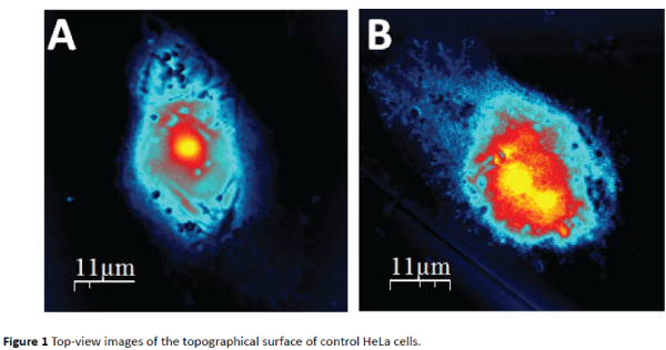
Figure 1: Top-view images of the topographical surface of control HeLa cells.
9578