Omer Engin1*,Oguzhan Sunamak2,Mebrure Evnur Uyar3 and Enis Unluturk1
1Buca Seyfi Demirsoy State Hospital,Surgery Department, Izmir, Turkey
2Haydarpasa Training and Research Hospital, Surgery Department, Istanbul,Turkey
3Buca Seyfi Demirsoy,Emergency Department, Izmir, Turkey
*Corresponding Author:
Omer Engin
Buca Seyfi Demirsoy State Hospital
Surgery Department
Izmir,Turkey
E-mail: omerengin@hotmail.com
Received: February 06, 2015; Accepted: January 12, 2016; Published: January 20,2016;
In our case presenting with complaints of sudden abdominal pain, nausea, and vomiting, abdominal tenderness and air under the diaphragm on the right were detected in direct abdominal graphy and diagnosed with peptic ulcers perforation, the patient was interned. As the clinical picture of ulcers perforation was incomplete in the follow-up, the tests were repeated and Chilaiditi could be diagnosed as the second direct graphy revealed a colon segment and haustras on the right under the diaphragm.
Introduction
Chilaiditi’s sign is an asymptomatic radiological finding that refers to the interposition of the colon between the liver and the diaphragm. Chilaiditi’s syndrome refers to the presence of concurrent symptoms such as abdominal pain, vomiting, constipation, and respiratory distress. Our case is presented here as he presented to us as a syndrome with symptoms and the entire colon was not detected initially and assumed to be free air and thus, surgical operation was planned due to the misdiagnosis of peptic ulcers perforation.
Case Presentation
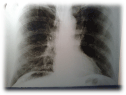
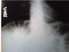
Our case is a 49-year-old male patient. He presented to our emergency clinic with complaints of sudden abdominal pain, nausea, and vomiting and abdominal tenderness in examination and air under the diaphragm on the right in direct abdominal graphy were detected (Figure 1). There was no leucocytosis. With current clinical findings, the patient was prediagnosed with peptic ulcers perforation and was interned. He was then followedup to allow for the complete clinical picture of peptic ulcer to form. As he developed no abdominal rigidity and leucocytosis in laboratory follow-up and his ultrasonography revealed no free abdominal fluid, his direct abdominal graphy was repeated and as a result colon segment and haustras on the right under the diaphragm were observed (Figure 2), the diagnosis of Chilaiditi’s syndrome replaced that of peptic ulcers perforation.

Figure 1: Visualization of free air under the diaphragm on the right in the first examination.

Figure 2: Visualization of colon haustras under the diaphragm on the right in the second examination.
Discussion
Approximately 10% of pneumoperitoneum cases are not associated with hollow organ perforation. There are many imitators of pneumoperitoneum including subphrenic abscess, colon volvulus, Chilaiditi syndrome, and so on [1].
Chilaiditi’s sign occurs in 0.1-0.25% of the population and is asymptomatic. Chilaiditi’s syndrome is a very rare condition with clinical symptoms. Main symptoms include abdominal pain,
distension, nausea, vomiting, and constipation. Abdominal pain is quite severe and might be confused with acute abdominal syndrome. It may sometimes result in respiratory distress and cardiac arrhythmias. It might also resemble the picture of renal colic [2,3].
In our case, the presence of sudden abdominal pain, tenderness in abdominal examination, and air under the diaphragm, but the failure to detect the colon haustras in the first film led to a misinterpretation of Chilaiditi’s syndrome as peptic ulcer.
Treatment of Chilaiditi syndrome is conservative; the symptoms are eliminated with bed rest, nasogastric decompression, softliquid diet, liquid replacement, and enema. Surgical intervention is rarely indicated [4,5].
Conclusions
Pneumoperitoneum is not an indicator of acute surgical abdomen. Differantial diagnosis should be made. Laboratory signs is supported with clinical signs and symptoms. If the diagnosis is suspicious , the patient is taken in observation and repeated control.
8489
References
- Chen CK, Su YJ, Lai YC, Tsai W, Chang WH (2008) Gas forming bacterial peritonitis mimics hollow organ perforation. Am J Emerg Med 26: 838.
- Senturk E (2007) Hepatodiyafragmatik interpozisyon: Chilaiditi Belirtisi. Solunum Hastaliklari 18: 130-133.
- Angulo Cuesta J, Gonzales Zorraguino A, Unda Urzaiz M, Flores Corral N (1991) Chilaiditi syndrome in the differential diagnosis of renal colic. Arch Esp Urol 44: 300-301.
- Kayacetin E, Gok M, Karaaslan H (2004) One of the rare reasons of abdominal pain: Chiliaditi syndrome, report of two cases. Akademik Gastroenteroloji Dergisi 3: 110-112.
- Kosar A, Tezel C, Orki A, Kiral H, Urek S, et al. (2004) Hepatodiyafragmatik interpozisyon: Chiliaditi sendromu:Olgu Sunumu. Izmir Gogus Hastanesi Dergisi 18: 133-135.


