Flyer

Journal of Biomedical Sciences

  • ISSN: 2254-609X
  • Journal h-index: 18
  • Journal CiteScore: 4.95
  • Journal Impact Factor: 4.78
  • Average acceptance to publication time (5-7 days)
  • Average article processing time (30-45 days) Less than 5 volumes 30 days
    8 - 9 volumes 40 days
    10 and more volumes 45 days
Awards Nomination 20+ Million Readerbase
Indexed In
  • Genamics JournalSeek
  • China National Knowledge Infrastructure (CNKI)
  • Directory of Research Journal Indexing (DRJI)
  • OCLC- WorldCat
  • Euro Pub
  • Google Scholar
  • J-Gate
  • SHERPA ROMEO
  • Secret Search Engine Labs
  • International Committee of Medical Journal Editors (ICMJE)
Share This Page

Research Article - (2019) Volume 8, Issue 1

Chromosomal Microarray Analysis Uncovers Pathogenic Copy Number Variations in Unexplained Neurodevelopmental Disorders and Congenital Anomalies

Saurabh Kumar Bhattacharya*, Leena Rawal, Shiba Ranjan Mishra, Richa Malhotra, Sumit and Vandana Lal

National Reference Laboratory, Department of Cytogenetics, Dr. Lal Path Labs Ltd., Block E, Sector 18, Rohini, 110085, Delhi, India

*Corresponding Author:

Saurabh Kumar Bhattacharya
National Reference Laboratory, Department of Cytogenetics, Dr. Lal Path Labs Ltd., Block E, Sector 18, Rohini, 110085, Delhi, India
Tel: +911130244157
E-mail: saurabhbhattacharya84@yahoo.in

Received date: March 14, 2016; Accepted date: April 19, 2016; Published date: April 22, 2016

Citation: Bhattacharya SK, Rawal L, Mishra SR, Malhotra R, Sumit, et al. (2019) Chromosomal Microarray Analysis Uncovers Pathogenic Copy Number Variations in Unexplained Neurodevelopmental Disorders and Congenital Anomalies. J Biomedical Sci Vol.8 No.1:3. doi: 10.4172/2254-609X.100099

Copyright: © 2019 Bhattacharya SK, et al. This is an open-access article distributed under the terms of the Creative Commons Attribution License, which permits unrestricted use, distribution, and reproduction in any medium, provided the original author and source are credited.

Visit for more related articles at Journal of Biomedical Sciences

Abstract

Background: Neurodevelopmental disorders represent a broad spectrum of cognitive, neurological, and/or psychiatric dysfunction caused by impairment of the brain during development. The present deals with the chromosomal rearrangements in patients with neurodevelopmental disorders, Developmental Delay/ Intellectual Disability (DD/ID) and/or congenital anomalies employing Chromosomal Microarray (CMA).

Methods: We used the CytoScan_750k array platform (Affymetrix) to analyze 102 patients with unexplained neurodevelopmental disorders and congenital anomalies. In this process, we have identified several deleted or duplicated genes possibly underlying the DD/ID phenotype to correlate the genetics with the clinical data.

Results: Of all the 102 patients identified with DD/ID, 48 patients had a normal profile (46XX/XY), 53 showed pathogenic CNVs along with an exceptional case (case 199), encompassing high levels of homozygosity (approx. 17.5%). The size of the CNVs in affected patients ranged from 36 kb to 15.5 MB. The most common variant in cases with ASD and developmental delay was duplication 22q11.2 of ~400Kb region, which was validated using karyotyping and FISH. Five out of 53 sporadic patients had known microdeletion syndromes. Case 66 showed 17p11.2 deletion; Smith-Magenis syndrome coupled with mosaic loss of chromosome 17p13.2, case 79 had a loss of Xp22.13 region overlapping with Rett syndrome, case 99 had 1q21.1 micro deletion syndrome along with Turner Syndrome, case 118 showed 4p16.3 terminal deletion; Wolf-Hirschhorn syndrome and case 122 had 7q11.23 deletion; William syndrome.

Conclusion: It is envisaged that the application of microarray will expand the spectrum of cytogenomic abnormalities by including complex and cryptic structural variants. Further, delineation of molecular mechanisms of these cytogenomic abnormalities coupled with the development of novel therapeutic approaches will ultimately lead to disease-specific personalized management and precision treatment.

Keywords

Neurodevelopmental disorders; Global developmental delay; Chromosomal microarray

Abbreviations

ASD: Autism Spectrum Disorder; CMA: Chromosomal Microarray; CNV: Copy Number Variation; ID: Intellectual Disability; GDD: Global Developmental Delay; KR: Mental Retardation

Introduction

The human brain is a highly complex structure, and its normal development and functioning are tightly orchestrated by a network of genes. The Neurodevelopmental Disorders (NDDs) are characterized by complex constellations of symptoms that include intellectual disability, emotional dysregulation and aberrant behaviors [1]. Sub microscopic chromosomal rearrangements have formed the foundation for neurodevelopmental disorders and are an important source of genetic and phenotypic variations. The prevalence rates of Neurodevelopmental Disorders (NDDs) from different regions of the country ranges from 1%-3% of the general population [2]. The various clinical entities such as Intellectual Disability (ID), Mental Retardation (MR), Global Developmental Delay (GDD), schizophrenia and Autism Spectrum Disorder (ASD), show considerable comorbidity and may be associated with a variety of neurological features within complex syndromes. Even with recent advances in genetic testing, there are still patients without an etiologic diagnosis, bearing the long diagnostic odyssey in search of accurate etiology. Some of these imbalances can be explained by gross chromosomal abnormalities, detected by conventional cytogenetic techniques such as GTG-banding. Although banding allows for the detection of numerical and structural chromosomal abnormalities present in the entire genome but have a limitation of resolution of 5-10 Mb and a detection rate of only 3%-5% [3]. Therefore, the etiology of congenital anomalies in 40%-60% of the cases remains unclear. In the past two decades, traditional banding has been combined with targeted molecular technologies to improve the resolution at which one can detect genomic changes.

The International Collaboration for Clinical Genomics (ICCG), also known as International Standard for Cytogenomic Array (ISCA) Consortium, has recommended CMA as the first-tier clinical diagnostic test for patients with DD, MR, and ASD of unknown causes [4]. Also, the American College of Medical Genetics and the American Academy of Paediatrics has endorsed, CNV analysis using array-based comparative genomic hybridization (Array-CGH) or CMA is now routinely performed in clinical genetics laboratories [5]. CMA detects Copy Number Variations (CNVs) in the entire genome with a much higher resolution than conventional cytogenetic. Recent studies using such genome-wide arrays to investigate patients with MR with and without dysmorphic features have suggested a diagnostic yield of 10%-25%, of which de novo findings count for approximately 10% [6-8]. The diagnostic yield of clinically significant CNVs varies between 12%-20%, depending on the clinical preselection and resolution of the array [9]. The CNVs can be divided into benign, pathogenic, likely pathogenic and CNVs of unknown clinical significance and can be polymorphic with a frequency greater than 1.0% or rare, less than 1.0%. The genomic disorders result from DNA rearrangements caused by de novo anomalies or are inherited, multiallelic or biallelic, Non- Allelic Homologous Recombination (NAHR) between regionspecific low copy repeats (LCRs) or non-homologous end joining, leading to interstitial deletions, duplications, and inversions as well as unbalanced translocations [10]. Our study is an attempt to unravel the application of microarray in patients with neurodevelopmental disorders in Indian Scenario. In the present study, we have used the CytoScan_750k array platform provided by the Affymetrix to analyze 102 patients with unexplained neurodevelopmental disorders and congenital anomalies to identify chromosomal rearrangements in these patients.

Methods

Clinical evaluation

A total of 102 patients with unexplained neurodevelopmental disorders and congenital anomalies were referred for chromosomal microarray analysis by physicians from pediatrics, rehabilitation, neurology, and psychiatric departments of different hospitals and research institutes. These patients were found to suffer from for unexplained DD, ID, MR, and ASD, microcephaly with or without dysmorphism or seizures. Written informed consent was obtained from patients or guardians for genetic analysis. The average age of patients ranged from 50 days to 32 years.

DNA preparation and chromosomal microarray

DNA was extracted from peripheral blood using DNAse blood and tissue kit (Qiagen) according to manufacturer's instructions and was stored at -200?C until further use. Genomic DNA concentration was measured by Nano drop spectrophotometer (Thermo Fisher). Approximately, 250 mg of high-quality genomic DNA was digested with Nsp1 restriction enzyme and digested DNA was then ligated to Nsp1 adapters. The ligation product was then amplified via Polymerase Chain Reaction (PCR) to produce amplicons in the range of 200 to 1100 bp. The amplicons are then purified and digested with DNAse I to produce 25 to 125 bp fragments. The fragments are end-labeled with a modified biotinylated base [11]. Samples were hybridized to Cytoscan 750k array in an Affymetrix Hybridization Oven at 600?C for 16 hours. Washes and staining of the arrays were performed with an Affymetrix Fluidics Station 450, and images were obtained using CytoScan 750K (Affymetrix, Santa Clara, CA, USA). The array is characterized with 750,000 CNV markers, including 200,000 genotype-able SNP probes and >550000 nonpolymorphism probes. The overall average marker space is 4125 base pairs. All data were visualized and analyzed with the Chromosome Analysis Suite (ChAS) software package (Affymetrix, ChAS V3.3) using Human Genome build hg19 and reporting threshold of the CNVs was set at 100 kb with a marker count of >50. The total number of autosomal, pseudo autosomal, intragenic and intragenic markers are 702,346; 811; 532,850 and 217,586, respectively.

Cytogenetic studies using G-banding standard karyotype at an average resolution of 750-800 bands and FISH analysis was also performed on the patients showing duplication of chromosome 22 at band q11.2. In order to perform FISH analysis, DiGeorge TUPLE1 (TUPLE1 [MIM 600237) (HIRA) region probe at 22q11.2 and a control probe, arylsulfatase-A (ARSA [MIM 607574]), at 22q13.3 (Vysis), were used on leukocytes in metaphase [12]. This is a direct-labeled dual-color probe mixture with TUPLE1 (HIRA) probe labeled in orange and ARSA probe labeled in green. Slide preparation, denaturation, hybridization, and posthybridization washes were all performed according to already established procedures and the manufacturer's recommendations, with minor modifications [12]. For each patient, at least 100 interphase and 10 metaphase cells were scored for both the TUPLE1 and ARSA signals.

Interpretation

Detected CNVs (gains/duplications or losses/deletions) were classified as pathogenic and likely pathogenic (possible clinical relevance), in accordance with the recommended guidelines from the International Standard Cytogenomic Array and the American College of Medical Genetics [13,14]. In this study, the pathogenic abnormalities has been classified as the ones where the detection of CNVs is in the known pathogenic regions, deletion/duplication are >3 Mb in size and deletions/ duplications <3 Mb and previously reported as pathogenic. Confidence is determined on a marker by marker basis by evaluating the concordance of the log ratio at each marker with the copy number state assigned by the hidden Markov model (HMM). The average confidence score of markers in gain and loss segments determines the confidence score of that segment.

The data were interpreted by using information available in the scientific literature, public databases and other general information about pathogenic CNVs (size, the content of Online Mendelian Inheritance in Man (OMIM) morbid genes or dosagesensitive genes, and type of dosage imbalance: duplication or deletion) [9]. Genomic map from the UCSC Genome Browser [15] was used to map the locations of CNVs and gene distribution. The Database of Genomic Variants [16] provided catalogues of structural variations found in normal healthy controls. The dbVar [17] database was also used to get information about CNVs from both normal and diseased populations. We also used the Database of Chromosomal Imbalance and Phenotype in Humans using Ensemble Resources [18] as a reference for known micro deletion and micro duplication syndromes, and the OMIM [19] for disease-causing genes, their functions and inheritance patterns.

Results

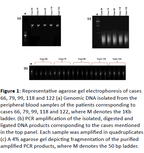
A total of 102 patients with GDD, ID, dysmorphism with or without multiple congenital anomalies were considered in the study. The genomic DNA was isolated, followed by digestion, ligation, PCR, fragmentation, and labeling (Figure 1). In this communication, only pathogenic or likely pathogenic CNVs have been reported. Out of 102 patients studied, 48 patients were found to have a normal karyotype of 46XX/XY. The pathogenic CNVs were found in 53 (57.6%) patients and an interesting case 199, showed the exclusive occurrence of high levels of homozygosity. The ratio of affected males to females was found to be (2.1:1). The rate of duplications was found to be more than that of deletions in the affected patients accounting to 74.3% and 25.6%, respectively.

jbiomeds-gel-electrophoresis

Figure 1: Representative agarose gel electrophoresis of cases 66, 79, 99, 118 and 122 (a) Genomic DNA isolated from the peripheral blood samples of the patients corresponding to cases 66, 79, 99, 118 and 122, where M denotes the 1Kb ladder. (b) PCR amplification of the isolated, digested and ligated DNA products corresponding to the cases mentioned in the top panel. Each sample was amplified in quadruplicates (c) A 4% agarose gel depicting fragmentation of the purified amplified PCR products, where M denotes the 50 bp ladder.

Patients with pathogenic CNVs

In 53 patients with pathogenic CNVs, 67.3% of patients had CNV involving single chromosome while 33.9% of patients showed co-occurrence of CNVs on two chromosomes.

Description of patients with pathogenic CNVs

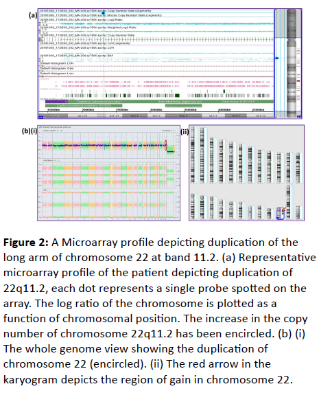
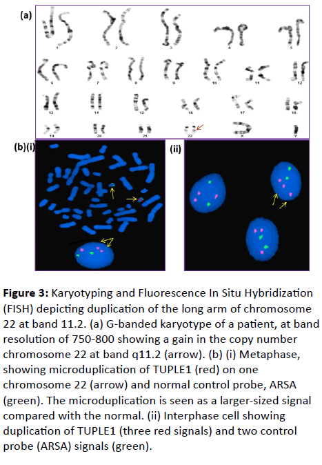
Of the total 53 patients, 18 (34.6%) showed duplication of 22q11.2 region detected by Cytoscan 750K microarray Figure 2. These duplications were later confirmed by karyotyping at a band resolution of 750-800 and by Fluorescence In Situ Hybridization (FISH) (Figure 3A and 3B). FISH analysis of interphase/metaphase cells revealed a duplication of the TUPLE1 probe on one chromosome 22q, reflecting increased gene dosage in the VCFS critical region (Figure 3B). A total of 100 cell nuclei were analyzed, and all cells had three red signals for TUPLE1 of similar intensity and two green signals of Locus- Specific Identifier (LSI) ARSA. The ARSA is the most commonly used commercial probe for internal control to determine the presence, deletion, or duplication of TUPLE1. The duplication was present in all 100 interphase cells screened and, duplication on chromosome 22 was visualized as a more intense signal of the TUPLE1 probe on one chromosome 22 as compared to its homolog in all cells (Figure 3B).

jbiomeds-Microarray-profile

Figure 2: A Microarray profile depicting duplication of the long arm of chromosome 22 at band 11.2. (a) Representative microarray profile of the patient depicting duplication of 22q11.2, each dot represents a single probe spotted on the array. The log ratio of the chromosome is plotted as a function of chromosomal position. The increase in the copy number of chromosome 22q11.2 has been encircled. (b) (i) The whole genome view showing the duplication of chromosome 22 (encircled). (ii) The red arrow in the karyogram depicts the region of gain in chromosome 22.

jbiomeds-long-arm

Figure 3: Karyotyping and Fluorescence In Situ Hybridization (FISH) depicting duplication of the long arm of chromosome 22 at band 11.2. (a) G-banded karyotype of a patient, at band resolution of 750-800 showing a gain in the copy number chromosome 22 at band q11.2 (arrow). (b) (i) Metaphase, showing microduplication of TUPLE1 (red) on one chromosome 22 (arrow) and normal control probe, ARSA (green). The microduplication is seen as a larger-sized signal compared with the normal. (ii) Interphase cell showing duplication of TUPLE1 (three red signals) and two control probe (ARSA) signals (green).

Patients with pathogenic CNVs and known syndromes

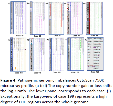
Five out of 53 sporadic patients (Case 66, 79, 99, 118 and 122) had known micro deletion syndromes (Figure 4). The Case 66 showed 17p11.2 deletion; Smith-Magenis syndrome coupled with the mosaic loss of chromosome 17p13.2, case 79 depicted loss of Xp22.13 region overlapping with Rett syndrome, case 99 had 1q21.1 micro deletion syndrome along with Turner Syndrome, case 118 showed 4p16.3 terminal deletion; Wolf- Hirschhorn syndrome and case 122 had 7q11.23 deletion; William syndrome. Four patients presented with previously reported but rare CNVs case 49 (22q13.33 and 22q11.2 duplication), case 56 (2q13, 4q35.1 and 15q11.2 duplication), case 115 (7p22.3 deletion and 10q25.3 duplication) and case 129 (15q13.3 and 17p13.3 duplication).

jbiomeds-copy-number

Figure 4: Pathogenic genomic imbalances CytoScan 750K microarray profile. (a to i) The copy number gain or loss shifts the log 2 ratio. The lower panel corresponds to each case. (j) Exceptionally, the karyoview of case 199 represents a high degree of LOH regions across the whole genome.

Micro duplication of 15q13.3 and 17p13.3

The case 129 showed micro duplication of both 15q13.3 and 17p13.3 regions (Figure 4). Individuals with duplication in 15q13.3 region showed variable symptoms like intellectual disability, communication difficulties, and behavioural problems including autism spectrum disorders [20]. Only one gene name Cholinergic receptor, Neuronal nicotinic, Alpha polypeptide 7 (CHRNA7) was found to be affected in the patient which is a critical gene associated with autism and behavioural disorders [21]. Also, micro duplication of 17p13.3 encompasses the same region that is deleted in Miller-Dieker syndrome [22]. Patients with 17p13.3 duplication exhibit neurobehavioral disorders, including delayed development, mental retardation, and attention deficit-hyperactivity disorder.

Mosaic loss of chromosome 17p13.2 and loss of 17p11.2

There was a loss of the short arm of the chromosome 17 at bands p13.2 and p11.2, respectively in case 66 (Figure 4). These regions were found to be 100% overlapping with the critical region of Smith-Magenis syndrome [23-25]. Smith-Magenis syndrome is associated with a deletion of the proximal short arm of chromosome 17, including the critical RAI1 gene region. Although the phenotype is variable, the syndrome can be suspected in patients with failure to thrive, brachycephaly (short head), prominent forehead, microcephaly (small head), flat and broad mid face, broad nasal bridge, strabismus, myopia, malformed ears, high and cleft palate, prognathism (protruding mandible), short and broad hands and feet, scoliosis (laterally curved spine), and cryptorchidism (undescended testes). Mental retardation is variable but usually severe with seizures and hyperactivity.

Micro deletion of chromosome Xp22.13

The case 79 had a clinical history of Global developmental delay with epileptic encephalopathy and dysmorphism. Microarray array analysis revealed a micro deletion of chromosome Xp22.13. This micro deletion resulted in a loss of five OMIM relevant genes (Figure 4). These genes included SCML2, CDKL5, RS1, PPEF1, and PHKA2. Out of all these five genes, CDKL5 is a main causative gene for epileptic encephalopathy which is an X-linked dominant severe neurological disorder characterized by the onset of seizures in the first months of life and severe global developmental delay resulting in mental retardation and poor motor control. Other features include lack of speech development, subtle dysmorphic facial features, sleep disturbances, gastrointestinal problems, and stereotypic hand movements. There is some phenotypic overlap with Rett syndrome [26,27].

Micro deletion of 7p22.3 and duplication of 10q25.3

A de novo micro deletion of chromosome 7p22.3 and duplication of chromosome 10q25.3 was identified in a 3-yearold boy (case 115) with loss of 2.5 Mb of chromosome 7p22.3 deletion and gain of 17.8 Mb of chromosome 10q25.3 (Figure 4). This abnormality corresponds to a der(7)t(7;10)(p22;q26) that is an unbalanced version of a t(7;10) translocation, which was paternally inherited. Investigation of the few genes in the region of chromosome 7p also revealed FAM20C and Platelet-Derived Growth Factor Alpha (PDGFA), as genes that have potential roles in patient's developmental delay and bone findings [28]. In case of, 10q25.3 duplication several putative candidate genes on 10q including DPYSL4 (neuronal differentiation), PPP2R2D (embryonic growth and development), INPP5A (intracellular signaling), and GPR123 (G-protein coupled receptor expressed in brain) may contribute to the pathogenesis in this patient [29-31].

Wolf-Hirschhorn syndrome (terminal deletion of 4p16.3)

A patient (case 118) was found to have ~ 6 Mb deletions at 4p16.3 region (Figure 4). The 4p deletion also known as Wolf- Hirschhorn syndrome came to existence in 1965 by Wolf et al, 1965 and Hirschhorn et al, 1965 [32,33]. It is a contiguous gene syndrome caused by partial loss of chromosomal region from the terminal region of chromosome 4p [34]. These patients are reported to have hypertelorism, arched eyebrows, epicanthic folds, short philtrum, micrognathia, hypotonia, seizures, EEG abnormalities and heart defects [35]. Patient 118 has similar phenotype such as Global developmental delay, failure to thrive and atrial septal defect. This genetic disorder is microdeletion syndrome with CNV of a variable size known to be caused by dosage-sensitive genes, and atypical recognized syndromes associated with non-recurrent micro deletions that might have been clinically missed at birth. Furthermore, even in a welldefined syndrome, non-recurrent chromosome deletions can be of different sizes, leading to a broad phenotypic spectrum.

The case with high LOH content

A case 199, corresponding to a female depicting dysmorphism, low set ears, and poor coordination showed extended contiguous regions of allele homozygosity (>8 Mb) in multiple chromosomes that is consistent with common descent (related parents). Several large regions of homozygosity were detected, encompassing approximately 17.5% of the genome. These may be added to provide a measure of identity by descent which in this case is equivalent to brother-half-sister parentage.

Discussion

Several neurodegenerative and neurodevelopmental disorders are now known to be caused by disparate recurrent and nonrecurrent genomic rearrangements that are mediated or stimulated by complex regional genomic architecture occurring throughout the human genome. The clinical utility of microarray technologies used in the post and prenatal diagnosis lies in its ability to detect sub microscopic copy number changes that are associated with clinically significant outcomes. Though the usefulness of CMA in identifying CNVs has been well recognized over the last decade but the data on its application in Indian scenario has not been explored. CMA analysis when conducted on 102 unexplained DD, MR and ASD patients, revealed duplication 22q11.2 region as the most frequent abnormality. The clinical phenotype of patients with this abnormality ranges from a mild learning disability to the presence of severe congenital malformations. The chromosome 22q11.2 region is highly susceptible to both micro deletions and duplications due to its misalignment of eight Low Copy Repeats (LCR) regions, LCR22-A through LCR22-H, which mediate nonallelic homologous recombination. Both micro deletions and micro duplications might be expected to occur at the same frequency. The micro duplication of chromosome 22q11.2 was faintly visible at higher resolution 750-800 in all the patients studied. Since 750-800 band resolution karyotypes are not routinely analyzed, FISH studies on interphase nuclei play a key role in the identification of patients with dup(22)(q11.2). Interphase FISH detected the duplication in all patients, but it was confirmed by metaphase FISH showing three TUPLE1 signals. Micro duplications of 22q11.2 have previously been diagnosed primarily using interphase FISH [36,37].

Pathogenic CNVs were found to be randomly distributed across the genome and were located on 1q21.1, 1p36, 4q13, 4q21, 7q11, 10q11.2, 15q11, 15q15.3, 22q13, Xq11, Xp22.13 and Xq27 regions of chromosomes (Table 1). Kaminsky et al. found that other than the common micro deletion syndromes, recurrent CNVs found were deletions in 16p13.11, 17q21.31, 17q12, 8p23.1 and 3q29 region, while recurrent duplications were found in 1q21.1, 16p13.11, 17q12, 22q11.2 regions [33]. In our study, no such recurrent deletions or duplications were found which could be attributed to a small number of cases. The size of pathogenic CNVs ranged between 36 kb to 15.5 Mb. This is similar to reported data in literature where these have been found to vary from 0.14 Mb to 17.58 Mb [38]. Among all the patients with pathogenic CNVs, thirty-five patients (67.3%) harbored a CNV on single chromosomes; whereas seventeen patients (33.9%) had CNVs on two different chromosomes. Patients in the latter category showed more prominent features for deletion, suggesting haploinsufficiency of genes had a more pronounced effect which leads to decreased protein levels and is less tolerated than duplication. Startlingly, high levels homozygosity in multiple chromosomes corresponding to case 199 validates the phenomenon that multiple generations of consanguinity can increase the levels of allele homozygosity. Although this result is not diagnostic of a specific condition, it raises the possibility of a recessive disorder with a causative gene located within one of these regions. Additionally, these results could indicate a familial relationship (first or second degree) between this individual's parents.

S.No. Case No. Age/Sex Clinical characteristics/Phenotype Copy No. Aberration Affected Chromosome Chromosome band/Cytoband Size (Kb) Number of OMIM affected Genes Molecular Karyotype/Genome Coordinates Classification
1 1 3Y/M Mild autism spectrum disorder, Speech delay, Poor eye contact, ADHD 0 Loss 1 q44 317 1 arr [hg19] 1q44 (246,174,603-246,491,736) × 1 Pathogenic
2 3 2/F Autism spectrum disorder, Speech delay, Hyperactive, Slightly delayed milestones, Sensory issues 3 Gain 6 q11.1 1037 1 arr [hg19] 6q11.1(61,886,426-62,923,825) × 3 Pathogenic
3 4 4/F Global developmental delay, facial dysmorphism             arr (1-22) × 2, (XX) × 1 Normal female
4 5 1Y/M Developmental delay with seizures 3 Gain 22 q11.22 484 2 arr [hg19] 22q11.22 (22,817,622-23,301,460) × 3 Pathogenic
5 6 7Y/M ADHD with autism spectrum disorder             arr (1-22) × 2, (XY) × 1 Normal male
6 7 6Y/M Behavioural disorder, facial dysmorphism with ADHD             arr (1-22) × 2, (XY) × 1 Normal male
7 8 13Y/M Autism, Delayed milestones, Facial asymmetry, Supernumerary teeth, No eye contact 1 Loss 1 q21.1 124 2 arr [hg19] 1q21.1(144,950,048-145,074,541) ×1 Likely pathogenic
        3 Gain 22 q11.22 330 1 arr [hg19] 22q11.22 (22,929,364-23,258,939) × 3 Likely pathogenic
8 9 5Y/M Behavioural issues 3 Gain 10 q11.22 853 3 arr [hg19] 10q11.22 (46,293,590-47,147,021) × 3 Likely pathogenic
9 10 3Y/M Developmental delay, Repetitive behaviour, Balance problem             arr (1-22) × 2, (XY) × 1 Normal male
10 11 2Y/F Gross motor delay, Hypotonia, Imbalance while walking, Dysmorphic facial features, Epicanthal folds 3 Gain 12 p13.31 500 7 arr [hg19] 12p13.31 (7,917,870-8,417,898) × 3 Likely pathogenic
11 12 3Y/M Global developmental delay, ADHD 3 Gain X p22.33 331 3 arr [hg19] Xp22.33  (313,456-644,440 or 263,456-94,440) × 3 Likely pathogenic
12 13 10Y/M Weakness, ? Developmental delay             arr (1-22) × 2, (XY) × 1 Normal male
13 14 4Y/M Dysmorphic facial features and developmental delay 3 Gain 13 q12.12 810 9 arr [hg19] 13q12.12q12.13 (25,446,646-25,736,308) × 3 Likely pathogenic
        2 Gain X q27.1 609 1 arr [hg19] 13q12.12q12.13 (25,446,646-25,736,308) × 3 Likely pathogenic
14 15 3Y/M Speech delay 3 Gain 22 q11.22 377 2 arr [hg19] 22q11.22 (22,901,370-23,258,939) × 3 Pathogenic
15 17 7M/M Multiple congenital anomalies, ASD?, B/L Retinoblastoma             arr (1-22) × 2, (XY) × 1 Normal male
16 18 33Y/F Risk of Down syndrome             arr (1-22) × 2, (XX) × 1 Normal female
17 20 6Y/F Failure to thrive, Speech delay, Gross motor delay, Intellectual disability, Increased Hypotonia, Seizures, Micrognathia, Retrognathia 3 Gain 22 q11.22 358 6 arr [hg19] 22q11.22 (22,901,370-23,258,939) × 3 Pathogenic
18 21 3Y/M Global Developmental Delay, ? fragile X syndrome, Intellectual disability 3 Gain 22 q11.22 358 6 arr [hg19] 22q11.22 (22,901,370-23,258,939) × 3 Pathogenic
19 22 11Y/F Behavioural issues, Speech Delay, Learning Disability, Intellectual Disability, Autism, Dimorphism 3 Gain 22 q11.22 377 2 arr [hg19] 22q11.22 (22,901,370-23,278,835) × 3 Pathogenic
20 23 4/F ? Rett syndrome, Autism, Speech delay, Motor delay 3 Gain 22 q11.22 400 3 arr [hg19] 22q11.22 (22,901,370-23,301,460) × 3 Pathogenic
21 28 2Y/F Developmental delay (motor and sensory) and stronger anxiety 3 Gain 22 q11.22 358 2 arr [hg19] 22q11.22 (22,901,370-23,258,939) × 3 Pathogenic
22 31 1 Y/M Global developmental delay             arr (1-22) × 2, (XY) × 1 Normal Male
23 32 1Y and 10 M/M Global developmental delay with hypotonia 3 Gain 22 q11.22 372 2 arr [hg19] 22q11.22 (22,929,364-23,301,460) × 3 Pathogenic
24 33 9 Years/M Hyperactive, Seizures, Short hands and Hypogonadism 3 Gain 1 p36.33 67 1 arr [hg19] 1p36.33 (2,174,958-2,242,417) × 3 Pathogenic
        3 Gain 16 p13.3 160 8 arr[hg19] 16p13.3 (2,053,327-2,213,040) × 3 Pathogenic
        3 Gain 19 p13.3 937 35 arr [hg19] 19p13.3 (675,955-1,612,855) × 3 Pathogenic
        3 Gain 22 q11.22 372 1 arr [hg19] 22q11.22 (22,929,364-23,301,460) × 3 Pathogenic
        2 Gain X q13.1 62 3 arr [hg19] Xq13.1 (70,481,039-70,542,994) × 2 Pathogenic
25 35 5 Y/M Autism Spectrum Disorder with ADHD 1 Loss 7 p21.3 172 1 arr [hg19] 7p21.3 (8,614,447-8,786,729) × 1 Pathogenic
        3 Gain 22 q11.22 372 2 arr [hg19] 22q11.22 (22,929,364-23,301,460) × 3 Pathogenic
26 36 4Y/F Behavioural issues, dysplastic toes and developmental delay 3 Gain 10 q11.22 3   arr [hg19] 10q11.22 (46,293,590-48,220,168) × 3 Pathogenic
        3 Gain 22 q11.22 2   arr [hg19] 22q11.22 (22,901,370-23,259,033) × 3 Pathogenic
        4 Gain X Q26.2 1   arr [hg19] 22q11.22(22,901,370-23,259,033) × 3 Likely Pathogenic
27 37 6Y/M Obesity, Delayed development, Slow learner, EEG anomaly and ?Prader Willi Syndrome 3 Gain 20 q13.33 199 7 arr [hg19] 20q13.33 (62,624,900-62,824,265) × 3 Likely Pathogenic
28 38 4Y/M Syndromic, Hyperactive, Impulsive, Speech delay 1 Loss 16 p12.2 775 3 arr [hg19] 22q11.22 (22,901,370-23,259,033) × 3 Pathogenic
29 39 3 Years/F Developmental delay, Hypotonia, Dysmorphic features and Micrognathia 3 Gain 4 q35.2 435 2 arr [hg19] 4q35.2 (190,522,320-90,957,460) × 3 Pathogenic
        3 Gain 22 q11.22 372 1 arr [hg19] 22q11.22 (22,929,364-23,301,460) × 3 Pathogenic
30 41 45 Months/F Delayed Milestones 3 Gain 22 q11.22 341 1 arr [hg19] 22q11.22 (22,927,618-23,268,533) × 3 Pathogenic
31 43 3Years/M Autism Spectrum Disorder 3 Gain 1 q11.22 22 2 arr [hg19] 22q11.22 (22,901,370-23,258,939) × 3 Pathogenic
32 44 3 Months/M Dysmorphic features, PDA syndrome and Radial Ray Anomaly 3 Gain 22 q11.22 358 2 arr [hg19] 22q11.22 (22,901,370-23,258,939) × 3 Pathogenic
33 45 9 Months/F Dysmorphic facies, Small mouth, Rocker bottom feet 3 Gain 3 q22.33 611 3 arr [hg19] 3q22.3q23 (138,215,113-38,825,892) × 3 Likely Pathogenic
        1 Loss 10 q11.22 1336 3 arr [hg19] 10q11.22 (46,989,206-48,203,688) × 3 Likely Pathogenic
        4 Gain 18 p11.32 18399 47 arr [hg19] 18p11.32q11.1 (136,227-18,534,784) × 4 Pathogenic
        3 Gain 22 q11.22 358 2 arr [hg19] 22q11.22 (22,901,370-23,258,939) × 3 Likely Pathogenic
        3 Gain X q28 75 4 arr [hg19] Xq28 (152,927,530-153,002,877) × 3 Likely Pathogenic
34 46 3 Years/ M Autism 3 Gain 22 q11.22 358 2 arr [hg19] 22q11.22 (22,927,618-23,258,939) × 3 Pathogenic
35 49 50 days/M Perinatal Hypoxia and Hypertonia 3 Gain 22 q11.22 331 1 arr [hg19] 22q11.22 (22,927,618-23,258,939) × 3 Pathogenic
        3 Gain 22 q13.33 36 1 arr [hg19] 22q13.33 (51,092,246-51,127,901) × 3 Pathogenic
36 57 15 years/m Convulsion and poor mental performance 3 Gain 22 q11.22 400 2 arr [hg19] 22q11.22 (22,901,370-23,301,460) × 3 Pathogenic
37 58 02Y/M Delayed Milestones 3 Gain 22 q11.22 377 2 arr [hg19] 22q11.22 (22,901,370-23,278,835) × 3 Pathogenic
38 59 02Y/M Autism, Speech delay and Hyperactivity 3 Gain 22 q11.22 400 2 arr [hg19] 22q11.22 (22,901,370-23,301,460) × 3 Pathogenic
39 60 03Y/M Macrocephaly and mild cortical artophy 3 Gain 22 q11.22 377 2 arr [hg19] 22q11.22 (22,901,370-23,278,835) × 3 Pathogenic
40 61 9 m/M Macrocephaly and mild cortical artophy 3 Gain 10 q11.22 1874 3 arr [hg19] 10q11.22 (46,293,590-48,167,553) × 4 Pathogenic
        3 Gain 17 p13.3 236 3 arr [hg19] 17p13.3 (811,480-1,047,339) ×  3 Pathogenic
41 62 5Y/m Autism Spectrum Disorder and Hyperactive 1 Loss 1 q21.1 121 2 arr [hg19] 1q21.1 (144,953,098-145,074,541) × 1 Pathogenic
        3 Gain 22 q11.22 330 1 arr [hg19] 22q11.22 (22,929,364-23,258,939) × 3 Pathogenic
42 68 5Y/m ? Speech delay             arr (1-22) × 2, (XY) × 1 Normal Male
43 69 7 Y/m Learning disability, Unable to understand simple things 3 Gain 22 q11.22 330 1 arr [hg19] 22q11.22 (22,929,364-23,258,939) × 3 Pathogenic
44 72 32m/M Behavioural problem             arr (1-22) × 2, (XY) × 1 Normal Male
45 74 4 Y/m Walking difficulty, developmental delay             arr (1-22) × 2, (XY) × 1 Normal Male
46 76 7 m/m Developmental delay, Hypotonia, Dysmorphic features             arr (1-22) × 2, (XY) × 1 Normal Male
47 77 6 Y/M Global developmental delay, Dysmorphic features             arr (1-22) × 2, (XX) × 1 Normal male
48 79 2 Y/F Global developmental delay with Epileptic encephalopathy and Dysmorphism 1 Loss X p22.13 596 5 arr [hg19] Xp22.13(18,346,842-18,943,282) × 1 Pathogenic
49 88 10Y/M ? Facioscapulohumeral dystrophy, ? Myotonic muscular dystrophy             arr (1-22) × 2, (XY) × 1 Normal male
50 95 27 m/M Speech delay, Gross motor delay, Learning disability, Autism             arr (1-22) × 2, (XY) × 1 Normal male
51 98 6Y/M sm spectrum disorder, Developmental delay             arr (1-22) × 2, (XY) × 1 Normal male
52 99 11 m/F Failure to thrive, IUGR 1 Loss 1 q21.1 906 8 arr [hg19] 1q21.1q21.2 (146,555,708-147,462,093) ×  1 Pathogenic
        1 Loss X p22.33 155065 711 arr [hg19] Xp22.33q28 (168,551-155,233,098) × 1 Pathogenic
53 100 15 m/M Facial dysmorphism, Broad nasal bridge, B/L Microtia, Right facial palsy             arr (1-22) × 2, (XY) × 1 Normal male
54 104 4Y/M Developmental delay             arr (1-22) × 2, (XY) × 1 Normal male
55 105 4Y/M MRI brain shows pansinusitis and adenoid hypertrophy 3 Gain 10 q11.12 2019 5 arr [hg19] 10q11.22 (46,252,072-48,270,746) × 3 Likely pathogenic
56 108 9y/M History of consanguineous marriage of parents, febrile seizures, ? visual problem, loss of bowel 3 Gain 22 q11.22 400 3 arr [hg19] 22q11.22 (22,901,370-23,301,460) × 3 Pathogenic
        3 Gain 22 q11.22 400 3 arr [hg19] 22q11.22 (22,901,370-23,301,460) × 3 Pathogenic
57 147 16 months/M Global developmental delay and Hypotonia             arr (1-22) × 2, (XY) × 1 Normal male
58 143 1y/M Dysmorphic features, low-level mosaic supernumerary marker chromosome detected in karyotype             arr (1-22) × 2, (XY) × 1 Normal male
59 154 2y/M ADHD with facial dysmorphism             arr (1-22) × 2, (XX) × 1 Normal male
60 136 11Y/F Periodic fever             arr (1-22) × 2, (XX) × 1 Normal female
61 139 3y/M Autism spectrum disorder 1 Loss 15 q15.3 109 4 arr [hg19] 15q15.3 (43,868,570-43,977,181) × 1 Likely pathogenic
62 141 7Y/F Developmental delay, short stature             arr (1-22) × 2, (XX) × 1 Normal female
63 137 2 months/F Severe pneumonia with dysmorphic facies 3 Gain 13 q14.11 73647 173 arr [hg19] 13q14.11q34 (41,460,947-115,107,733) × 3 Pathogenic
64 129 32Y/F Autism spectrum disorder 3 Gain 15 q13.3 444 1 Arr [hg19] 15q13.3 (31,999,631-32,444,043) × 3 Pathogenic
        3 Gain 17 p13.3 211 3 arr [hg19] 17p13.3 (716,837-927,465) × 3 Pathogenic
65 117 19 months/M Developmental delay             arr (1-22) × 2, (XY) × 1 Normal male
66 115 3y/M Developmental delay, GDD with paternally inherited unbalanced t(7;10) 1 Loss 7 p22.33 2570 23 arr [hg19] 7p22.3 (43,376-2,613,293)  × 1 Pathogenic
        3 Gain 10 q25.3 17841 79 arr [hg19] 10q25.3q26.3(117,585,175-135,426,386) × 3 Pathogenic
67 138 4y/M Developmental delay, autistic features, seizures, dysmorphic course features             arr (1-22) × 2, (XY) × 1 Normal male
68 116 11y/M Assessment of language and hearing skill             arr (1-22) × 2, (XY) × 1 Normal male
69 119 3y/F Delayed development milestone             arr (1-22) × 2, (XX) × 1 Normal female
70 118 10 m/M Global developmental delay, Failure to thrive, Atrial Septal Defect 1 Loss 4 p16.3 4589 47 arr [hg19] 4p16.3p16.2 (68,345-4,656,856) × 1 Pathogenic
        1.48 Loss mosaic 4 p16.3 6368 55 arr [hg19] 4p16.3p16.1 (68,345-6,435,945) × 1-2 Pathogenic
71 122 8 m/M Microcephaly, Strabismus, Failure to thrive 1 Loss 7 q11.23 1596 27 arr [hg19] 7q11.23 (72,621,345-74,217,791) × 1 Pathogenic
72 125 22y/M Intellectual disability             arr (1-22) × 2, (XY) × 1 Normal male
73 155 14y/F Developmental delay, difficulty in walking, mild cerebellar atrophy 2.2 Gain mosaic X p22.31 14203 67 arr [hg19] Xp22.31p22.12 (6,056,862-20,259,724) × 2-3 Pathogenic
74 140 15y/M Seizures, Epilepsy             arr (1-22) × 2, (XY) × 1 Normal male
75 159 3y/M To rule out any chromosome abnormalities             arr (1-22) × 2, (XY) × 1 Normal male
76 152 2y/M Autism spectrum disorder             arr (1-22) × 2, (XY) ×1 Normal male
77 173 7y/F Developmental delay             arr (1-22) × 2, (XX) × 1 Normal female
78 175 2y/M Microcephaly and Spasticity 1.52 Loss mosaic 22 q11.1 16417 165 arr [hg19] 22q11.1q12.3(16,888,899-33,305,441) × 1-2 Pathogenic
        1 Loss 22 q21.1 3152 44 arr [hg19] 22q11.21 (18,648,855-21,800,471) × 1 Pathogenic
79 176 6y/M Facial Dysmorphism             arr (1-22) × 2, (XY) × 1 Normal male
80 177 4M/M ?Leri weill dyschondrosteosis             arr (1-22) × 2, (XY) × 1 Normal male
81 179 2y/M Autism 3 Gain 22 q11.22 468 3 arr [hg19]  22q11.22 (22,833,258-23,301,460) × 3 Pathogenic
82 180 15y/M Low IQ and Abnormal Facies 2 Gain X q11.2 7072 39 arr [hg19]  Xq11.2q13.1(63,706,926-70,779,158) × 2 Pathogenic
83 181 14y/M Low IQ and Abnormal Facies 2 Gain X q11.2 7072 39 arr [hg19]  Xq11.2q13.1 (63,706,926-70,779,158) × 2 Pathogenic
84 182 8y/M Intellectual Disability 2 Gain X q11.2 7072 39 arr [hg19]  Xq11.2q13.1 (63,706,926-70,779,158) ×2 Pathogenic
85 186 11y/F Hearing disability and behaviour concern             arr (1-22) × 2, (XX) × 1 Normal female
86 188 3y/M Autism Spectrum Disorder             arr (1-22) × 2, (XY) × 1 Normal male
87 197 08 Y/ m Learning disability and speech delay             arr (1-22) ×2, (XY) ×1 Normal male
88 198 20Y/F Sclerosis             arr (1-22) × 2, (XX) × 1 Normal female
89 199 10 Y/F Dysmorphism, Low set ears and poor coordination             arr (1-22) ×2, (XX) ×1 High Long Continuous Regions of Homozygosity brother-half-sister parentage
90 205 1Y/F Developmental delay, dysmorphic features and poor growth 1.39 Loss mosaic 14 q32.13 12,197 95 arr [hg19]  14q32.13q32.33 (95,087,352-107,284,437) × 1-2 Pathogenic
        1 Loss 14 q32.2 5,022 40 arr [hg19]  14q32.2q32.31 (97,163,311-102,185,159) ×  1 Pathogenic
91 209 2Y/M Dysmorphic facial features, hypotonia and failure to thrive 3 Gain 22 q11.22 358 2 arr [hg19]  22q11.22 (22,901,370-23,258,939) × 3 Pathogenic
92 66 06Y/F Global developmental delay 1.73 Loss Mosaic 17 p13.2 29211 248 arr [hg19]  17p13.2q12 (5,239,141-34,450,123) × 1-2 Pathogenic
        1 Loss 17 p11.2 3688 35 arr [hg19]  17p11.2 (16,745,600-20,433,723) × 1 Pathogenic
93 210 01Y/M Global developmental delay             arr (1-22) × 2, (XY) × 1 Normal Male
94 211 03Y/M Autism             arr (1-22) × 2, (XY) × 1 Normal Male
95 212 05Y/M Intellectual Disability             arr (1-22) × 2, (XY) × 1 Normal Male
96 213 07Y/M Delayed development milestone             arr (1-22) × 2, (XY) × 1 Normal Male
97 214 03Y/M Autism, Speech delay and Hyperactivity             arr (1-22) × 2, (XY) × 1 Normal Male
98 215 01Y/M Developmental delay             arr (1-22) × 2, (XY) × 1 Normal Male
99 216 07Y/M Delayed Milestones             arr (1-22) × 2, (XY) × 1 Normal Male
100 217 09Y/M autism spectrum disorder             arr (1-22) × 2, (XY) × 1 Normal Male
101 218 02Y/F Intellectual Disability             arr (1-22) × 2, (XX) × 1 Normal female
102 219 04Y/F Global developmental delay             arr (1-22) × 2, (XX) × 1 Normal female

Table 1: Clinical features and cytogenetic findings of patients with pathogenic CNVs.

Thus, high-resolution screening using microarray not only detects sub microscopic chromosomal imbalances but also allows accurate delineation of the duplicated or deleted chromosomal segment. Our data involving detailed phenotype analysis and CNVs can add to databases for future genetic studies for discovering new candidate genes and molecular pathways underlying unexplained neurodevelopmental disorders.

Conclusion

Our study is an attempt towards deconvoluting the effect of copy number variants in genetic diseases and more broadly in the clinical evaluation of patients with unexplained DD/ID, congenital anomalies and dysmorphic features. Further, enhancements in genomic microarray analysis will soon allow the reliable analysis of all copy number variations throughout the chromosome at the kilobase or single exon resolution. Also, clarification of the genetic profile generated by CMA coupled with knowledge-based genetic counseling, rational clinical action and follow up familial studies may aid in directing medical management.

Acknowledgment

Authors are grateful to the patient and their family to support the case study. We would also like to thank the management of Dr. Lal Path Labs for providing excellent facilities.

Conflict of Interest

The author(s) declare they have no conflict of interests.

24220

References

  1. Lee JA, Lupski JR (2006) Genomic rearrangements and gene copy-number alterations as a cause of nervous system disorders. Neuron 52: 103-121.
  2. Arora NK, Nair MKC, Gulati S, Deshmukh V, Mohapatra A, et al. (2018) Neurodevelopmental disorders in children aged 2-9 years: Population-based burden estimates across five regions in India. PLoS Med 15: e1002615.
  3. Shevell M, Ashwal S, Donley D, Flint J, Gingold M, et al. (2003) Practice parameter: evaluation of the child with global developmental delay: Report of the quality standards subcommittee of the american academy of neurology and the practice committee of the child neurology society. Neurology 60: 367-380.
  4. Yobb TM, Somerville MJ, Willatt L, Firth HV, Harrison K, et al. (2005) Microduplication and triplication of 22q11.2: A highly variable syndrome. Am J Hum Genet 76: 865-876.
  5. Schaefer GB, Mendelsohn NJ (2013) Clinical genetics evaluation in identifying the etiology of autism spectrum disorders: 2013 guideline revisions. Genet Med 15: 399-407.
  6. Vissers LE, de Vries BB, Osoegawa K, Janssen IM, Feuth T, et al. (2003) Array-based comparative genomic hybridization for the genomewide detection of submicroscopic chromosomal abnormalities. Am J Hum Genet 73: 1261-1270.
  7. Tyson C, Harvard C, Locker R, et al. (2005) Submicroscopic deletions and duplications in individuals with intellectual disability detected by array-CGH. Am J Med Genet A 139: 173-185.
  8. Rosenberg C, Knijnenburg J, Bakker E, Friedman JM, Langlois S, et al. (2006) Array-CGH detection of micro rearrangements in mentally retarded individuals: clinical significance of imbalances present both in affected children and normal parents. J Med Genet 43: 180-186.
  9. Miller DT, Adam MP, Aradhya S, Biesecker LG, Brothman AR, et al. (2010) Consensus statement: Chromosomal microarray is a first-tier clinical diagnostic test for individuals with developmental disabilities or congenital anomalies. Am J Hum Genet 86: 749-764.
  10. Alaerts M, Del-Favero J (2009) Searching genetic risk factors for schizophrenia and bipolar disorder: Learn from the past and back to the future. Hum Mutat 30: 1139-1152.
  11. Slater HR, Bailey DK, Ren H, Cao M, Bell K, et al. (2005) High-resolution identification of chromosomal abnormalities using oligonucleotide arrays containing 116,204 SNPs. Am J Hum Genet 77: 709-726.
  12. Crifasi PA, Michels VV, Driscoll DJ, Jalal SM, Dewald GW (1995) DNA fluorescent probes for diagnosis of velocardiofacial and related syndromes. Mayo Clin Proc 70: 1148-1153.
  13. Kearney HM, Thorland EC, Brown KK, Quintero-Rivera F, South ST (2011) American College of Medical Genetics standards and guidelines for interpretation and reporting of postnatal constitutional copy number variants. Genet Med 13: 680-685.
  14. Richards S, Aziz N, Bale S, Bick D, Das S, et al. (2015) Standards and guidelines for the interpretation of sequence variants: a joint consensus recommendation of the American College of Medical Genetics and Genomics and the Association for Molecular Pathology. Genet Med 17: 405-424.
  15. Beal JC (2014) Case report: Neuronal migration disorder associated with chromosome 15q13.3 duplication in a boy with autism and seizures. J Child Neurol 29: NP186-188.
  16. van Bon BW, Mefford HC, Menten B, Koolen DA, Sharp AJ, et al. (2009) Further delineation of the 15q13 microdeletion and duplication syndromes: a clinical spectrum varying from non-pathogenic to a severe outcome. J Med Genet 46: 511-523.
  17. Roos L, Jonch AE, Kjaergaard S, Taudorf K, Simonsen H, et al. (2009) A new microduplication syndrome encompassing the region of the Miller-Dieker (17p13 deletion) syndrome. J Med Genet 46: 703-710.
  18. Potocki L, Chen KS, Park SS, Osterholm DE, Withers MA, et al. (2000) Molecular mechanism for duplication 17p11.2-the homologous recombination reciprocal of the Smith-Magenis microdeletion. Nat Genet 24: 84-87.
  19. Smith ACM, Boyd KE, Elsea SH, Finucane BM, Haas-Givler B, et al. (2001) Smith-Magenis Syndrome. In: Pagon RA, Adam MP, Ardinger HH, et al. editors. Gene Reviews (Internet). Seattle (WA): University of Washington, Seattle, 2001 (Last update 2012): 1993-2014.
  20. Elsea SH, Girirajan S (2008) Smith-Magenis syndrome. Eur J Hum Genet 16: 412-421.
  21. Mei D, Marini C, Novara F, Bernardina BD, Granata T, et al. (2010) Xp22.3 genomic deletions involving the CDKL5 gene in girls with early onset epileptic encephalopathy. Epilepsia 51: 647-654.
  22. Fehr S, Wilson M, Downs J, Williams S, Murgia A, et al. (2013) The CDKL5 disorder is an independent clinical entity associated with early-onset encephalopathy. Eur J Hum Genet 21: 266-273.
  23. Cuvertino S, Stuart HM, Chandler KE, Roberts NA, Armstrong R, et al. (2017) ACTB Loss-of-function mutations result in a pleiotropic developmental disorder. Am J Hum Genet 101: 1021-1033.
  24. Balikova I, Menten B, de Ravel T, Le Caignec C, Thienpont B, et al. (2007) Subtelomeric imbalances in phenotypically normal individuals. Hum Mutat 28: 958-967.
  25. Balikova I, Vermeesch JR, Fryns JP, Van Esch H (2009) Bronchiectasis and immune deficiency in an adult patient with deletion 2q37 due to an unbalanced translocation t(2;10). Eur J Med Genet 52: 260-261.
  26. Minhas HM, Pescosolido MF, Schwede M, Piasecka J, Gaitanis J, et al. (2013) An unbalanced translocation involving loss of 10q26.2 and gain of 11q25 in a pedigree with autism spectrum disorder and cerebellar juvenile pilocytic astrocytoma. Am J Med Genet A 161: 787-791.
  27. Wolf U, Reinwein H, Porsch R, Schroter R, Baitsch H (1965) Deficiency on the short arms of a chromosome No. 4. Humangenetik 1: 397-413.
  28. Hirschhorn K, Cooper HL, Firschein IL (1965) Deletion of short arms of chromosome 4-5 in a child with defects of midline fusion. Humangenetik 1: 479-482.
  29. Battaglia A, Carey JC, Wright TJ, (2001) Wolf-Hirschhorn (4p-) syndrome. Adv Pediatr 48: 75-113.
  30. Battaglia A, Filippi T, Carey JC (2008) Update on the clinical features and natural history of Wolf-Hirschhorn (4p-) syndrome: Experience with 87 patients and recommendations for routine health supervision. Am J Med Genet C Semin Med Genet 148C: 246-251.
  31. Edelmann L, Pandita RK, Spiteri E, Funke B, Goldberg R, et al. (1999) A common molecular basis for rearrangement disorders on chromosome 22q11. Hum Mol Genet 8: 1157-1167.
  32. Ensenauer RE, Adeyinka A, Flynn HC, Michels VV, Lindor NM, et al. (2003) Microduplication 22q11.2, an emerging syndrome: Clinical, cytogenetic, and molecular analysis of thirteen patients. Am J Hum Genet 73: 1027-1040.
  33. Kaminsky EB, Kaul V, Paschall J, Church DM, Bunke B, et al. (2011) An evidence-based approach to establish the functional and clinical significance of copy number variants in intellectual and developmental disabilities. Genet Med 13: 777-784.
  34. Siggberg L, Ala-Mello S, Jaakkola E, Kuusinen E, Schuit R, et al. (2010) Array CGH in molecular diagnosis of mental retardation-A study of 150 Finnish patients. Am J Med Genet A 152: 1398-410.