Case Report - (2022) Volume 16, Issue 2
Cilium Incarnatum Externum and Its Management-A Rare Presentation
Dr. Seemantini Ayachit1* and
Dr. Khaled Amin Helaiwa2
1Chief Registrar, Department of
Ophthalmology BDF Hospital, Bahrain
2Consultant, Department of Ophthalmology BDF Hospital, Bahrain
*Correspondence:
Dr. Seemantini Ayachit,
Chief Registrar, Department of
Ophthalmology BDF Hospital,
Bahrain,
Tel: 97339412869,
Email:
Received: 29-Jan-2022, Manuscript No. iphsj-22-12405;
Editor assigned: 31-Jan-2022, Pre QC No. Preqc No.12405;
Reviewed: 14-Feb-2022, QC No. QC No.12405;
Revised: 19-Feb-2022, Manuscript No. iphsj-22-12405(R);
Published:
28-Feb-2022, DOI: 10.36648/1791-809X.16.2.914
Abstract
A 39 years old male presented to our clinic with complains of a small swelling and redness over his right upper eyelid with mild pain, but no discharge. On examination, an aberrant eyelash was seen growing subcutaneously under the swollen area. This aberrant eyelash was removed surgically.
Introduction
Cilia Incarnata means misdirection of eyelashes. It has two major types:
• Cilium Incarnatum Externum: in which the eyelash grows in the subcutaneous plane [1-5].
• Cilium Incarnatum Internum: in which the eyelash grows subconjunctivally [1,2, 6].
Both conditions are rare. Herein we present a case of Cilium Incarnatum extrenum and its management.
Case Report
A 39 years old male presented to our clinic with complains of a small swelling on his right upper eyelid with mild pain, irritation, redness and mild itching over it. He had no visual symptoms, no discharge and there was no history of ocular trauma or chronic eye rubbing.
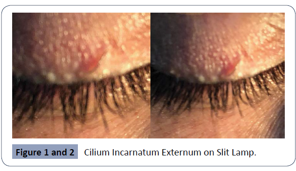
On slit lamp examination an obliquely placed eyelash was seen running subcutaneously just above the right eye upper lid margin. All other eyelashes were normally directed. [Figures A&B]. A clinical diagnosis of Cilium Incarnatum Externum was made and a surgical removal was planned.
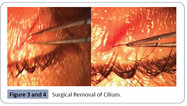
The aberrant eyelash over the right upper eyelid was removed after making a small superficial incision over the lesion and the eyelash was pulled out with the help of a fine non-tooth forceps. The procedure was performed under topical anaesthesia under slit lamp bi-microscope. [Figures &D] The cilium was about 4 mm in length and was thinner than the normal eyelashes. Tobradex eye ointment was prescribed for local application twice daily for a period of five days (Figure 1-4).
Figure 1 and 2 Cilium Incarnatum Externum on Slit Lamp.
Figure 3 and 4 Surgical Removal of Cilium.
Discussion
Cilia Incarnata or misdirected eyelashes can either be found running subcutaneously- Cilium Incarnatum Externum or Subconjunctivally- Cilium Incarnatum Internum [1-4].
Both conditions are considered hereditary [2,6]. The patient with Cilium Incarnatum Externum [3,4] usually presents either with cosmetic concern due to a bump over the eyelid margin or due to scratchy irritation over the lesion causing discomfort to the patient. It is a rare benign condition in which the whole eyelash along with its follicle is misdirected. To best of our knowledge very few cases have been reported so far, the last one was reported in May 2021
Eyelashes are ectodermal in origin and both Cilium Incarnatum Externum and Internum are uncommon and under reported. The under reporting could be due to the minimum symptoms that these conditions cause and due to less attention to the lid margin on slit lamp bimicroscope. Also these cases can be congenital as well as acquired [1, 4].
In cilium Incarnatum Externum, the treatment involves incision over the eyelid just above the buried eyelash followed by its removal by simple epilation; we adopted the same method in our case [2-4].
Differential diagnosis includes Trichiasis, Distichiasis [7]. Tristichia &Tetrastichiasis polystichia [8]
Trichiasis is defined as a normal eyelash growing inwards and the problem is only in the direction of the eyelashes. It is usually seen secondary to Trachoma, Chronic Blepharitis, chemical burns, Meibomian gland dysfunction, Ocular cicatricial pemphigoid and Stevens-Johnson syndrome.
Distichiasis is a condition where there is separate row of eyelashes behind the normal row of eyelashes. Primary Distichiasis is a congenital condition and can be Autosomal dominant as seen in association with Lymphedema- Distichiasis syndrome. Secondary causes are same as those of Trichiasis.
Tristichia and Tetrasticia are rare conditions where the patients have three or four rows of eyelashes respectively [9]
Ectopic cilia are very rare in humans. They are a congenital disturbance of the position of the eyelashes, which are usually on the lateral quadrant of the upper eyelid or conjunctival surface of the eyelid [2].
Conclusion
Cilia Incarnata can be easily managed if correct diagnosis is made. They are detected under high magnification on a slit lamp bimicroscope. If a patient presents with a lesion on outer lid margin; a diagnosis of Cilium Incarnatum Externum should be kept in mind. We presented this case as it is both an unusual and under reported condition.
REFERENCES
- Cilia Incarnata, Bhupendra C. Patel , Raman Malhotra (2021) Cilia Incarnata that Pearls Treasure Island (FL): Stat Pearls Publishing. PMID: 30725664.
Indexed at, Google scholar, Crossref
- Prathiba D, Nivean, Ramesh Dorairajan, varshini Ramesh, Nivean Madhivanan (2021) Cilia Incarnatum: A rare Variant of an eye lash. Ophthalmic Image 1:447.
Google scholar, Crossref
- Dongargaonkar, surendra, Preeti A patil (2020) Cilium Incarnatum Externum-Suahrut -Ophthalmic Image year 68:204.
Indexed at, Google Scholar, Crossref
- Taru Dewan, Chetna Sharma, Harshik Chawla (2007) Cilium Incarnatum Extrenum: Digit J Ophthalmol 23:16-17.
Indexed at, Google scholar, Crossref
- F Gutteridge (2002) Curious Cilia cases. Clin Exp Optomary 85:306-308.
Indexed at, Googlescholar, Crossref
- HS Aggarwal, WK Munshi (1966) Aberrant Eyelashes. IJO 14:253-257.
Indexed at, Google Scholar
- Sen D.K, hariMohan D, Gupta K (1996) Cilium Inver sum: Brit J Ophthal 9:207.
Indexed at, Google scholar, Crossref
- Agarwal H S (1963) Cilium Inversum. Am J Ophthalmol 55:648-649.
Indexed at, Google Scholar, Crossref
- Richard Johnson (2006) clinical and experimental optometry an unusual case of an extra row of eye lashes in the superior eyelid skin. 89:322-324.
Indexed at, Google scholar, Crossref
Citation: Citation: Ayachit S, Helaiwa KA, et al. (2022) Cilium Incarnatum Externum and Its Management – A Rare Presentation. Health Sci J. Vol. 16 No. 2: 914.


