Ayush Dubey*
Department of Neurology, SAIMS Medical College and PG Institute, Indore, Madhya Pradesh, India
*Corresponding Author:
Ayush Dubey
Department of Neurology
SAIMS Medical College and PG Institute
Indore, Madhya Pradesh, India
Tel: +917999867326
E-mail: ayushdubey2@yahoo.co.in
Received Date: April 03, 2018; Accepted Date: April 20, 2018; Published Date: April 24, 2018
Citation: Dubey A (2018) CT Negative SDH. Ann Clin Lab Res Vol.6: No.2:233.
Description
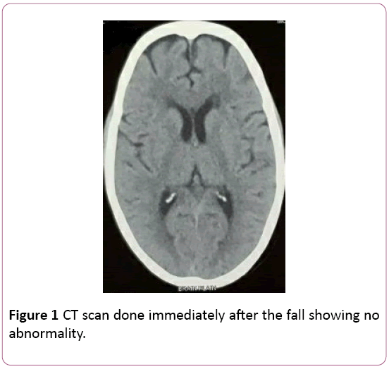
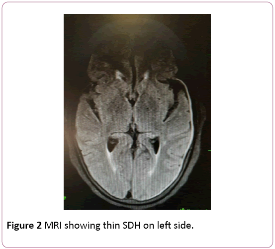
CT scan is the gold standard diagnostic investigation for head injuries as it is a rapid investigation, easily available and cost effective even though MRI is a more sensitive and accurate tool [1]. However, small hematomas, parenchymal injuries or vault fractures near the bone may be missed on CT. Thin SDH being close to the bone might be missed on CT scan and similarly, SAH can also go undetected on CT scan (Figures 1 and 2). The same reason explains the drawbacks of CT in detecting small traumatic lesions of the brain stem or posterior fossa structures [2,3].

Figure 1: CT scan done immediately after the fall showing no abnormality.

Figure 2: MRI showing thin SDH on left side.
Although being a sensitive tool, a negative CT scan does not always rule out acute SDH and emphasis should be to find out the hemorrhage either by thinner cuts of CT or by doing MRI in all such suspected cases wherever possible.
22480
References
- Vinchon M, Noule N, Tchofo PJ, Soto-Ares G, Fourier C, et al. (2004) Imaging of head injuries in infants: temporal correlates and forensic implications for the diagnosis of child abuse. J Neurosurg 101: 44-52.
- Ashis P,D Singh, Khandelwal N (2006) Fallacies of routine CT scans in identifying lesions in severe head injury. Ind J Neurotrauma 3: 36-40.
- Weber C, Grzyska U, Lehner E, Adam G (2003) Clinical relevance of cranial CT under emergency conditions—basic neuroradiologic investigations. Rofo 175:654-662.


