AMC Kumarihami1, SDL Heshani1, Sathyathas Puvanasunthararajah1* and Rasika Illeperuma2
1Department of Radiography and Radiotherapy, General Sir John Kotelawala Defence University, Sri Lanka
2Department of Medical Laboratory Science, University of Peradeniya, Sri Lanka
*Corresponding Author:
Sathyathas Puvanasunthararajah
Faculty of Allied Health Sciences
Department of Radiography and Radiotherapy
General Sir John Kotelawala Defence University, Sri Lanka
Tel: 779552885
E-mail: pthas88@gmail.com; ckumarihami@yahoo.com
Received Date: 02 February 2018; Accepted Date: 20 February 2018; Published Date: 27 February 2018
Citation: Kumarihami AMC, Heshani SDL, Puvanasunthararajah S, Illeperuma R (2018) Development of Brief Image Quality Evaluation Criteria for Digital OrthoPantomography (OPG) Images in Dental Radiography for Sri Lanka. Health Sci J. Vol. 12 No. 1: 551.
Copyright: © 2018 Kumarihami AMC, et al. This is an open-access article distributed under the terms of the creative commons attribution license, which permits unrestricted use, distribution and reproduction in any medium, provided the original author and source are credited.
Keywords
Orthopantomography; Dental radiography; Evaluation criteria
Introduction
Dental radiography is the art of recording images of a patient’s oral structures by using x rays. There are two main methods in imaging the oral structures according to the place of the films; intra oral radiography film is placed inside of the mouth and extra oral radiography film is placed outside of the mouth. Dental panoramic radiography is one of the methods of extra oral radiography.
Dental panoramic radiography is a unique extra oral film technique that allows the dentist to view the entire dentition and related structures [1]. Quality assurance of dental panoramic radiographs is very important as properly planned quality control tests and quality management programs contribute in producing a good quality image. Good quality image is the basic means to proper diagnosis.
Dental panoramic radiography imaging is mostly used for orthodontic assessments. Therefore image quality should not be minimized to avoid misinterpretation. In panoramic imaging both principal of tomography and principal of scanning is used [2]. Hence correct positioning of the dental arch inside the focal trough is important to obtain images with high diagnostically value. Images can be obtained as plain film radiographs and digital radiographs.
There are different ways and methods to assess the quality of an Ortho Pantomography (OPG) images. Around the world in different clinical setups and with different equipment facilities, many researchers have assessed the quality of the OPG films and many of them have observed the occurrence of large number of errors throughout the process of the production of panoramic radiographs [3]. According to the depth of our knowledge there only few researches have done on quality evaluation of panoramic images in Sri Lanka.
This study main purpose was to develop brief image quality evaluation criteria for orthopantomography (OPG) in dental radiography for radiographers. Each factor in quality evaluation criteria contributes in different proportion to the overall image quality. The study focused to zoom out common errors related to OPG images and the results can be used to minimize those possible errors. Minimizing image repetition directly affects in reducing patient dose [4]. Also time reduction of both patients’ and the hospital cannot be neglected as time is the best source of money.
Objectives of the study
Develop brief image quality evaluation criteria accordance with the established criteria for digital orthopantomography (OPG) to identify the most frequent errors.
Materials and Methods
This was a retrospective cross sectional study. The approval obtained from administrative boards of respective hospitals and ethical review committee of General Sir John Kotelawela Defence University. The study represented all the digital OPG images that have been taken from January to December in 2015 from one government and private hospital.
Sampling and sample size
Systematic sampling method was used to select digital OPG images. According to the population ratio 75 of digital images were selected from the government hospital and 25 of digital images selected from the private hospital.
Method of image evaluation
Pretest was conducted among six OPG images. In these study three BSc radiographers with more than two years’ experience of working only on dental radiography and three four year undergraduate students of General Sir John Kotelawela Defence University (two male students and two female students) selected randomly for image evaluation. Each image was given to the evaluators and asked to observe according to the data collection tool. Adequate time was given to access each image (Table 1).
Table 1 Main categories and sub categories of the evaluation tool.
| Category |
Sub categories |
| Category 01 (Identification) |
Name, age, sex, date, registration number and anatomical marker |
| Category 02(Artifact) |
foreign body compromise the anatomical area and Motion artifact |
| Category 03(Anatomical coverage) |
Top infra orbit exclusion, bottom margin isnot at the lower border of the mandible and TM joint is not clearly seen |
| Category 04 (Patient positioning) |
Tongue is not positioned against the palate, bite block in not visualizing, lips are not closed, anterior teeth positioning error, incorrect Frankfort plane positioning, head rotated, head tilted, patient is in slumped position (Not stand in ski position), lead apron or thyroid collar positioned too high, motion artifact |
Equipment and material
The Owandy I- max touch 3D machine which can perform digital panoramic imaging and 3D cone beam CT was used. Features of the equipment are 220-240 V with 50/60 Hz and maximum exposure time is 14 seconds, resolution is 92 μm and dimension of images size is 130 × 130 mm, with 512 × 512 pixels.
Statistical analysis
Minitab version 14 used to data analyze with 0.05 significant levels with 95% confidence level. Descriptive statistics, parametric and non-parametric tests were used appropriately. Paired t-test and Kruskal Wallis tests were used. Validation of the criteria has tested by following two methods.
Validation of the criteria is assessed by frequent errors according to evaluators’ response: Average of the responses given by each evaluator and evaluator category (radiographer and student radiographer) were compared for main and sub categories of the data collection tool to validate the criteria, then the calculated values used to see if there was any significant difference in observation between each evaluator and evaluator’ category. Strongly deviated evaluators were excluded and selected evaluator’ final results were given. If the majority gave same answer, it is suggested criteria was effective and easily understandable.
Validation of the criteria by the overall marks by the weighted marks: The data collection tool was given to selected senior experience radiographers and asked to weight the sub and main categories. According to the results marks were finalized (Table 1). Those marks were used for parametric and non-parametric statistical analysis. The image quality was categorized into three groups (excellent quality (Over 80%), average quality (between 50%-80%) and low quality (below 50%). Then the outcome of every image was compared among the seven evaluators.
Results
Image quality response comparison among radiographers and radiography students
Kruskal Wallis used as a non-parametric test. There were no significant differences between the responses of the evaluators (p>0.05). Average overall quality marks for radiographers and student radiographers were calculated. The results showed that the both distributions are normal distributions while both were dependent variables. Therefore paired t-test was used to compare the variables. There was no significant difference (p<0.05) between the responses of the radiographers and student radiographers (Figure 1).
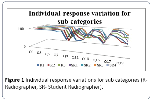
Figure 1: Individual response variations for sub categories (RRadiographer, SR- Student Radiographer).
The most frequent error by each evaluator’s responses were tongue is not against the palate reported by three evaluators, top infra orbit excluded, lips not closed and Bite block is not visualizing said by two and one evaluators respectively. Results of the six evaluators showed the most frequent error was anatomical coverage from category 03 (Table 2).
Table 2 Overall responses on frequent errors of sub categories according to category.
| Category |
Questions |
| Category 01 (Identification) |
All are same frequently occurring |
| Category 02(Artifact) |
Foreign body compromise the anatomical area, motion artifact |
| Category 03(Anatomical coverage) |
Top infra orbit exclusion, TM joint not clearly seen and bottom margin not at the lower border of the mandible |
| Category 04 (Patient positioning) |
Tongue is not positioned against the palate, bite block in not visualizing, lips not closed, anterior teeth positioning error, incorrect Frankfort plane positioning, head rotated head tilted, patient is in slumped position (Not stand in ski position) andlead apron or thyroid collar positioned too high |
Category 1: Identification acquired not only the highest mark but also the complete mark. Hence there is no identification errors found in the sample. Category 2: Artifact acquired 97% value therefore it has a low frequent appearance. But it is the 3rd most frequent error of the sample. Category 3: Anatomical coverage gained the lowest points of 73.3% according to the evaluation. Therefore it was the highest frequent error category of the data sample. Category 4: Patient positioning acquired 77.7% and it is the 2nd most frequent error of the sample (Table 3).
Table 3 Individual responses on frequent error (sub category wise) (R- Radiographer, SR- Student Radiographer). Table 4 Overall image quality assessments.
| Evaluators |
The most frequent error |
Second most frequent error |
| R1 |
Tongue is not against palate 54% |
Top infra orbit excluded 57% |
| R2 |
Top infra orbit excluded 44% |
Tongue is not against palate 48% |
| R3 |
Top infra orbit excluded 50% |
Tongue is not against palate 51% |
| SR 1 |
Lips not closed 50% |
Tongue is not against palate53% |
| SR 2 |
Tongue is not against palate 48% |
Top infra orbit excluded |
| SR 3 |
Bite block is not visualizing 39% |
Tongue is not against palate 49% |
| SR 4 |
Tongue is not against palate 38% |
Bite block is not visualizing 47% |
Overall responses on image quality assessment
According to the marks on the weighted chart, an overall mark is given for each image for each individual. Excellent quality was above 80%, average quality and poor quality was above 50%-80% and below 50% respectively (Table 4).
Table 4 Overall image quality assessments.
| Sameresponses (Excellent, Average or low quality) fromthe evaluators |
| Ratio |
07-Jul |
06-Jul |
05-Jul |
04-Jul |
| Percentage |
57 |
18 |
10 |
14 |
Discussion
The study accessed the validity of the data collection tool by comparing the evaluation responses of each evaluator. There was no evidence of already established standard criteria for digital OPG images in Sri Lanka. A study has done in Sri Lanka to develop an inventory by comparing it to modified Korean criteria (MKC) [1], consequently it was vital to develop criteria which was practicable in Sri Lankan practices. According to the results overall image quality values have no significant difference between the evaluation of radiographers and student radiographers. Also there was no significance different between each evaluator (p>0.05), When considering average value of the responses were given by seven evaluators. It gives the highest frequent error and the least frequent error. The highest mean value gives the least frequent error and the lowest mean value gives the highest frequent error.
Responses of the individual evaluation for each sub category of the data collection tool showed few sub categories as the most frequent error for each evaluator. Three of the evaluators’ responses showed that the most frequent error was absence of tongue against palate. Responses of radiographer 1 (R1) was 54%, student radiographer 2 (SR2) was 48% and student radiographer 3 (SR3) was 48% for the absence of tongue against the palate was the most frequent error. Also responses of radiographer 2 (SR2) was 48%, radiographer 3 was 51% student radiographer 1 (SR1) was 53% and student radiographer 3 (SR3) was 49% for the absence of tongue against the palate was the second most frequent error. The results values were not widely deviated from each other. Similar study on investigation of the most common positioning error showed the absence of tongue against palate as the most frequent error [5]. When considering the average value for all the seven evaluators’ the most frequent error was absence of positioning the tongue against the palate (41.28%). The possible causes of absence of positioning the tongue against the palate became as the most frequent error may be due to failure to give proper instruction by the radiographer or failure to follow the instructions. The main disadvantage is that radiographer can’t observe the positioning of tongue hence it may be hard to correct it in many circumstances.
Category of identification showed as the least frequent error of the images and it received total marks. Digital OPG examination required certain patient details to implement the examination. Therefore ignorance of patient details has a low possibility. According to the responses, the least frequent error category was anatomical coverage while the most frequent error was absent of positioning the tongue against the palate (41.28%) which was a component in patient positioning. Images were categorized into three categories, excellent quality, average quality and low quality images. A similar study has done for investigate image quality of OPG images [6]. The results of the study conveyed the quality outcome of each image was same for each other for 57% which is more than the half of the images [7-13].
Conclusion
According to the results the selected frequent error and overall image quality of radiographer to student radiographers or between evaluators doesn’t change significantly and the quality evaluation criteria may be an easily understandable and sophisticated one to be used.
22004
References
- Nileema A, Pallegama RW, Nuwan D, Jayasinghe RD (2016) Development of a brief inventory for quality evaluation of panoramic radiography. Sri Lanka Dent J 45: 55-62.
- Choi BR, Choi H, Huh KH, Yi WJ, Heo MS, et al. (2012) Clinical image quality evaluation for panoramic radiography in Korea dental clinics. Imaging Sci Dent 42: 183-190.
- Vano E, Guibelaide E, Morillo A, Pedrosa CS, Fernandez JM (1995) Evaluation of European image quality criteria for chest examinations. British J Radiol 68: 1349-1355.
- Dhillon M, Raju SM, Verma S, Tomar D, Mohan RS, et al. (2012) Positioning errors and quality assessment in panoramic radiography. Imaging Sci Dent 42: 207-212.
- Decusara M, Miliceseu V (2011) Image quality assessment of orthopantomograms, Romanian JOral Rehabilitation 3: 54-58.
- Gulsahi A (2011) Bone quality assessment for dental implants.In: Implant dentistry – The most promising discipline of dentistry(2ndedn.).nTech, Turkey. pp: 12-16.
- Kullman L, Joseph B (2006) Quality of digital panoramic radiography in a newly published dental school. Sweden Dent J 30: 165-170.
- Lannucci JM, Howerton LJ (2006) Dental radiography principles and techniques (3rd edn.). Elsevier, Missouri. pp:45-50.
- Offiah AC, Hall CM (2003) Evaluation of the commission of the European communities’ quality criteria for the paediatric lateral spine. British J Radiol 76: 885-890.
- Rondon RH, Pereira YC, Nasciment GC (2014) Common positioning errors in panoramic radiography:Review. Imaging Sci Dentistry 44: 1-6.
- Wahab HA, Ferguson DJ, Kheir NA (2016) Assessment of panoral radiograph quality in a dental treatment center. Research Gate 6: 85-94.
- Whaites E (2002) Essentials of dental radiography and radiology (3rdedn.).Churchil Livingstone, London. pp: 68-90.

