Keywords
|
| Intermittent ataxia; Ependymal cyst in pediatric population; Non-colloid cyst; Third ventricle; Ball valve mechanism; Obstructive hydrocephalus; Endoscopic ablation |
Introduction
|
| Ependymal cysts are rare, neuro-ectoderm derived, benign lesions; mostly diagnosed incidentally on MRI [1]. The most common location is lateral ventricle but they have also been identified in subarachnoid spaces and brain parenchyma [1]. We present a case of ependymal cyst in an 18 months old girl, presented with intermittent ataxia and vomiting. The cyst presented antero-superiorly in the third ventricle; a site common for endoderm derived, colloid cysts (0.5-1% of all brain tumors) but not for ependymal cysts. While ependymal cysts very rarely present in this age group, the unique location and dynamic behavior in our case lead to the intermittent clinical symptoms. Endoscopic cyst ablation resulted in excellent recovery and complete resolution of symptoms. |
Case Report
|
|
History
|
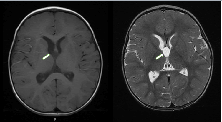
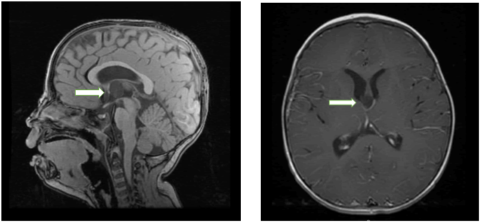
| An 18 month old Caucasian girl presented with episodic difficulty in walking, intermittent ataxia, broad-based gait and vomiting. Patient experienced three such episodes, over the course of three weeks. Other than the suggestive ataxia, neurologic examination including funduscopic examination was unremarkable. Initially, a diagnosis of acute labrythitis was considered but excluded following an ENT consultation. Following the third unexplained episode, a non-contrast head CT was ordered which revealed a mildly dilated right lateral ventricle and a third ventricular cyst that was isoattenuating to the CSF (cerebrospinal fluid) (Figure 1). MRI revealed mildly dilated lateral ventricles and an intraventricular cyst 1.7 x 1. 5 x 1.1 cm, located anteriorly in the third ventricle, at the level of Foramen of Monro (Figures 2 and 3). The child’s symptoms were considered secondary to the cyst occluding the foramen in a ball valve manner, explaining the episodic and paroxysmal nature of the presentation. |
|
Treatment
|
| Patient was admitted to the ICU (intensive care unit) and endoscopic ablation of the cyst was planned. Neurosurgical ablation was performed through a right frontal approach followed by endoscopic third ventriculostomy. |
|
Histology and immunophenotype
|
| The lining epithelium of this cyst consisted of simple cuboidal epithelial cells without goblet cells and no prominent cilia. The epithelial cells showed diffuse positivity for glial fibrillary acidic protein and dot-like positivity for epithelial membrane antigen. Histologic findings and the immunophenotype (negative cytokeratin, positive S100 and variable Glial fibrillary acidic protein immunoreactivity) were consistent with a diagnosis of ependymal cyst. |
|
Post-op Course
|
| Postoperative hospital course was uncomplicated. Complete resolution of symptoms was reported on one month follow-up visit. |
Discussion
|
| Third ventricle cysts are almost exclusively colloid and they usually present in 3rd to 5th decade of life [2]. Ependymal cysts hardly ever present in third ventricles, their most common location being lateral ventricles. Third ventricle cysts, although very rare in children, can present as chronic headaches, papilledema and diplopia [3]. The acute presentation can be in the form of nausea, vomiting, drop attacks and transient loss of consciousness secondary to acute hydrocephalus [4-7]. |
| Rarely, they have also been described as a sudden cause of death [8-10]. The anterosuperior pendulous attachment in the third ventricle can impart a mobile character to the cyst, leading to a ball effect resulting in intermittent obstruction of Foramen of Monro. The literature suggests that this mechanism is thought to explain the paroxysmal behavior and clinical symptoms. Of interest, the patient symptoms were not associated with abrupt movements of the head. Symptoms were primarily vomiting and ataxia which goes against Bruns syndrome. The imaging findings effectively rue out metabolic abnormalities, Bruns syndrome, non-specific syncope and bobble-head doll syndrome. |
| Since third ventricular cysts are usually colloid; they appear hyperdense on CT, hyperintense on T1 and hypointense on T2 weighted images. Signal intensity on neuroimaging depends on the amount of protein content in them [11]. In our case, imaging characteristics did not support a diagnosis of a colloid cyst. The cyst was considered not to be a colloid cyst as it appeared isoattenuating to the CSF on CT scan. On MRI there were abnormal signals identified on all sequences; appearing hypointense on T1, hyperintense on T2 with no diffusion restriction or contrast enhancement. |
| Asymptomatic cysts of should be monitored closely [12]. However, if cysts become symptomatic, surgical treatment should be considered. Among various microsurgical options available for third ventricular colloid cysts, the transcallosal approach is reported to have the most favorable outcome [13,14]. Recent studies show neuro-endoscopic ablation of the cyst to be superior to a microsurgical approach due to low infection risk and smaller number of patients requiring ventriculoperitoneal shunting [12,15], In our case, endoscopic fenestration of the cyst wall into the subarachnoid space lead to quick recovery and complete resolution of symptoms. |
Conclusion
|
| Although third ventricular cysts are extremely uncommon in infants and children, they should be considered in the differential diagnosis of intermittent symptoms that are suggestive of raised intracranial pressure. The knowledge of this potentially fatal cause of intermittent ICP in the pediatric population, can hasten the diagnosis. Early endoscopic ablation of the cyst is associated with full recovery and minimal morbidity. |
| This case in an 18 month old with an ependymal cyst presenting at the level of the foramen of Monro and mimicking signs and symptoms of a colloid cyst may represent the youngest child reported in the literature. |
Figures at a glance
|
 |
 |
 |
| Figure 1 |
Figure 2 |
Figure 3 |
|
| |
|
|

