Flyer

Archives in Cancer Research

  • ISSN: 2254-6081
  • Journal h-index: 14
  • Journal CiteScore: 3.77
  • Journal Impact Factor: 4.09
  • Average acceptance to publication time (5-7 days)
  • Average article processing time (30-45 days) Less than 5 volumes 30 days
    8 - 9 volumes 40 days
    10 and more volumes 45 days
Awards Nomination 20+ Million Readerbase
Indexed In
  • China National Knowledge Infrastructure (CNKI)
  • CiteFactor
  • OCLC- WorldCat
  • Publons
  • Geneva Foundation for Medical Education and Research
  • Euro Pub
  • Google Scholar
  • J-Gate
  • Secret Search Engine Labs
  • International Committee of Medical Journal Editors (ICMJE)
  • Zenodo
Share This Page

Image - (2015) Volume 3, Issue 3

Epithelioid Hemangioendothelioma of the Lumbar Spine

Lalya I1, Maghous A2,*, Marnouche E2 and Mansouri H3
  1. Radiation Oncologist; Department of radiotherapy, Mohamed V Military Hospital, Rabat, Morrocco
  2. Resident of Radiation Oncology; National Institute of Oncology, Rabat, Morrocco
  3. Associate Professor of Radiation Oncology; Department of radiotherapy, Mohamed V Military Hospital, Rabat, Morrocco
Corresponding Author: Abdelhak Maghous, Resident of Radiation Oncology, National Institute of Oncology, Rabat, Tel: 00212661572770, E-mail: magabdelhak@gmail.com
Related article at Pubmed, Scholar Google
Visit for more related articles at Archives in Cancer Research

Abstract

A 25-year-old man was referred for irradiation of a vertebral Epithelioid hemangioendothelioma (EHE) with a 6-month history of non systematized lombo-sciatic pain. There was no history of direct or indirect trauma or overexertion. At presentation the patient had severe back pain and difficulty walking. A neurological examination revealed percussion pain over the lumbar spine and cauda equina syndrome with perineal sensory symptoms and weakness of the muscles of the lower legs.

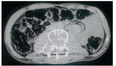
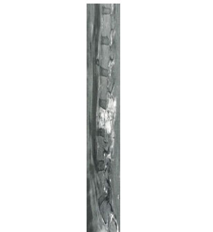
A 25-year-old man was referred for irradiation of a vertebral Epithelioid hemangioendothelioma (EHE) with a 6-month history of non systematized lombo-sciatic pain. There was no history of direct or indirect trauma or overexertion. At presentation the patient had severe back pain and difficulty walking. A neurological examination revealed percussion pain over the lumbar spine and cauda equina syndrome with perineal sensory symptoms and weakness of the muscles of the lower legs. The computed tomography (CT) revealed a radiolucent lesion of the L3 vertebral body which destroyed the cortex and extending into adjacent soft tissues (Figure 1). A lumbar magnetic resonance imaging confirmed the presence and the extent of the lesion accompanied by compressive epiduritis (Figure 2). To decompress the spinal cord, a laminectomy of the L3 vertebra with L2-L4 osteosynthesis was performed. A bone biopsy confirmed the diagnosis of an EHE after immunohistochemistry assays. Four weeks later and after three session of physiotherapy, distal flaccid paraplegia was permanent. The patient was then addressed to our department for definitive RT of her bone lesion. The total prescribed dose was 45 Gy in 25 fractions (1.8 Gy per fraction, once daily, 5 days a week). Actually, the patient is currently being treated.

Figures at a glance

Figure 1 Figure 2
Figure 1 Figure 2
7207