Keywords
|
| Punica granatum l.var spinosa; Malignant cell line; Cancer; Chemotherapy; Cytotoxicity; Anticancer effects |
INTRODUCTION
|
| Breast cancer is the most common of all cancers and is the leading cause of cancer deaths in women worldwide, accounting for >1.6% of deaths and case fatality rates are highest in low resource countries (www.tmc.gov.in). Unlike other cancers, breast cancer is eminently treatable if detected at an early stage. Although the incidence of breast cancer has increased globally over the last several decades (Hortobagyi, Anderson, Porter), the greatest increase has been in Asian countries (Green et al.) [1]. |
| More women in all risk categories are seeking information regarding their individual breast cancer risk, and there is a need for their primary care clinicians to be able to assess familial risk factors for breast cancer, provide individualized risk information, and offer surveillance recommendations. Estimates of the number of women with a family history of breast cancer range from approximately 5% to 20%, depending on the population surveyed. Many of these women will not have a family history that suggests the presence of a highly penetrant breast cancer susceptibility gene. However, a small subset of such women will come from families with a striking incidence of breast and other cancers often associated with inherited mutations. The development and refinement of risk prediction models provide an epidemiologic basis for counseling women with a family history that does not appear related to a dominant susceptibility gene. In contrast, the recent isolation of BRCA1, the localization of BRCA2, and the acknowledgment that additional breast cancer susceptibility genes must exist provide a molecular basis for counseling some high-risk women (Kent F. Hoskins et al.). |
| Chemotherapy is the treatment of cancer with one or more cytotoxic antineoplastic drugs (chemotherapeutic agents) as part of a standardized regimen. Among the most successful chemotherapeutic agent are Cyclophosphamide, methotrexate, 5-fluorouracil and Doxorubicin. All of these agents enhance serious side effects or long term complication (Corrie PG, Pippa G). These side effects include kidney damage, hearing loose, lower blood count, liver damage, nerve damage, and blood vessel damage (Laskova, Majtas, Oconnor, and Fitzpatrick). |
| A cell line refers to cells derived from a single cell type that have been adapted to grow continuously in the laboratory and are used in research (www.cancer.gov). The MCF-7 cell line is estrogen responsive and contains the estrogen receptor (AATC, May, 2008). MCF-7 is a cell line that was first isolated in 1970 from the breast tissue of a 69-year old Caucasian woman (https://www.mcf7.com/). MCF-7 is the acronym of Michigan Cancer Foundation-7, referring to the institute in Detroit where the cell line was established in 1973 by Herbert Soule and co-workers (Soule) The Michigan Cancer Foundation are now known as the Barbara Ann Karmanos Cancer Institute (www.cancer.gov). |
| On Thursday, January 2, 1997, Dr. Herbert Soule, the scientist who developed the MCF-7 breast cancer cell line, died. At the time, we were in the process of writing this tribute to mark the 25th anniversary of Dr. Soule's remarkable accomplishment. The cells, derived from a breast cancer patient in the Detroit area and developed at the Michigan Cancer Foundation, Detroit, became a standard model in hundreds of laboratories around the world. In retrospect, the story of the diverse uses of these cells is really the history of our developing knowledge of hormone-regulated cell replication, and they provided a unique insight into the endocrine therapy of breast cancer [2]. Our article is offered as a tribute and memorial to Dr. Soule. We will trace research with MCF-7 cells to illustrate the change in our ideas about cell replication and to highlight the advances in our understanding of the signal transduction pathway of estrogen and the molecular biology of estrogen action. All of these advances depended on the unique properties of MCF-7 cells. The cell system has now found applications in experimental therapeutics, and the results from these studies are being translated to the clinic for the treatment of patients (Anait S. Levenson and Craig Jordan). |
| Cytotoxicity can also be monitored using the 3-(4, 5-Dimethyl- 2-thiazolyl)-2, 5-diphenyl-2H-tetrazolium bromide (MTT) or MTS assay. Viable cells will reduce the MTS reagent to a colored form azan product. A similar redox-based assay has also been developed using the fluorescent dye, resazurin. In addition to using dyes to indicate the redox potential of cells in order to monitor their viability, researchers have developed assays that use ATP content as a marker of viability (Riss). Such ATP-based assays include bioluminescent assays in which ATP is the limiting reagent for the luciferase reaction (Fan and Wood). |
| The cell line has been widely studied and exhibits relatively slow growth characteristics. Both EPA and DHA have been shown to have inhibitory effects on MCF-7 cell proliferation. In other studies, Cellular Toxicity of Nanogenomedicine in MCF-7 Cell Line: MTT assay (Yadollah Omidi et al.) and Cytotoxicity of methanol extracts of Elaeis guineensis on MCF-7 (Soundararajan Vijayarathna, Sreenivasan Sasidharan) this cell line has been used [3]. |
|
Punica Granatum L. var spinosa
|
| The pomegranate /?p?m??ræn?t/, botanical name Punica granatum, is a fruit-bearing. The pomegranate is widely considered to have originated in the vicinity of Iran and has been cultivated since ancient times. Punica granatum has been claimed in traditional literature to be valuable against a wide variety of diseases, such as kidney stone, bleeding of kidney, irritable condition of bladder inflammation, painful urination, burning sensation, problem in urine discharge (Ahmad, Mehmood) (Figure 1). |
| The pharmacological mechanisms of pomegranate and the individual constituents responsible for them [4,5]. Extracts of all parts of the fruit appear to have therapeutic properties, and tree have medicinal benefit as well. Current research seems to indicate the most therapeutically beneficial pomegranate constituents are ellagic acid ellagitannins (including punicalagins), punicic acid, flavonoids, anthocyanidins, anthocyanins, and estrogen flavonols and flavones. The goal of many pomegranate studies has been to identify the therapeutic constituents (Koushan Sineh Sepehr et al.) (Figure 2). |
| The use of juice, peel, and oil has also been shown to possess anticancer activities, including interference with tumor cell proliferation, cell cycle, invasion, and angiogenesis [6]. The toxicity of Punica granatum L. var. spinosa has not been intensively studied. |
|
Therapeutic applications
|
| The pomegranate, Punica granatum L., is an ancient, mystical, unique fruit borne on a small, long-living tree cultivated throughout the Mediterranean region, as far north as the Himalayas, in Southeast Asia, and in California and Arizona in the United States. In addition to its ancient historical uses, pomegranate is used in several systems of medicine for a variety of ailments [7-9]. The synergistic action of the pomegranate constituents appears to be superior to that of single constituents. In the past decade, numerous studies on the antioxidant, anticarcinogenic, and anti-inflammatory properties of pomegranate constituents have been published, focusing on treatment and prevention of cancer, cardiovascular disease, diabetes, dental conditions, erectile dysfunction, bacterial infections and antibiotic resistance, and ultraviolet radiation-induced skin damage [10,11]. Other potential applications include infant brain ischemia, male infertility, Alzheimer's disease, arthritis, and obesity (Jurenka). |
| Pomegranate (Punica granatum L.) is known to possess pharmacological activities, such as antioxidant and anticancer [12-14]. In this study, we evaluated the antioxidant potency of a methanolic pomegranate fruit peel extract (PPE) and the relation with its antiproliferative and apoptotic effects on MCF-7human breast cancer cells [15,16] (Tables 1-3). |
Extraction process
|
|
Extraction procedure for Punica granatum L. var spinosa
|
|
Preparation of plant extract
|
| 1. The seeds and peels parts of the plant were separated, shade dried, and grinded into powder with mortar and pestle. |
| 2. The prepared powder was kept in tight containers protected completely from light. |
| 3. Extraction of ethanolic extract was carried out by macerating 100 g of powdered dry plant in 500 mL of 70% ethanol for 48 h at room temperature. |
| 4. Then, the macerated plant material was extracted with 70% ethanol solvent by percolator apparatus (2-liter volume) at room temperature. |
| 5. The plant extract was removed from percolator, filtered through Whatman filter paper (NO. 4), and dried under reduced pressure at 37°C with rotator evaporator. |
| 6. The ethanol extract was filtered and concentrated using a rotary evaporator and then evaporated to dryness. |
| 7. Briefly, the concentrated plant extracts were dissolved in dimethyl sulphoxide (DMSO) (SIGMA, USA) to get a stock solution of 10 mg/mL. |
| 8. The substock solution of 0.2 mg/mL was prepared by diluting 20 μL of the stock solution into 980 μL serum-free culture medium, DMEM (the percentage of DMSO in the experiment should not exceed 0.5) (Table 1 and Figure 3). |
|
Cell culture
|
| MCF-7 cells collected from Thapar University, Punjab, were used. Cells were maintained in monolayer culture in T75 flasks at 37°C in DMEM containing 100 μg/ml streptomycin and 10% fetal bovine serum (FBS) in an atmosphere of 95% air and 5% CO2. All cell culture work was carried out on an aseptic LAF-bench, and all equipment used in direct contact with cells was sterilized or autoclaved [17]. |
|
Passaging MCF-7 cells
|
| Cells were passaged when they were 90% confluent, about every fourth day. Media was removed by aspiration, and 3 ml of PBS were added to wash the media from the cells. To detach cells 1 ml of trypsin was added and cells were incubated at 37°C for 5 minutes. The culture plate was tapped gently against the palm to detach cells, and cells were checked to be detached under the microscope. Cells were transferred to a centrifuge tube by adding 10 ml DMEM, and centrifuged at 1250 rpm (Allegra 6R Centrifuge, Beckman) for 5 minutes. The supernatant was removed, and the tube was “flipped” to separate cells. Cells were resuspended in 10 ml of DMEM [18]. After the resuspension half of the cell stock was transferred into a culture plate containing 5 ml DMEM Media, PBS and trypsin was heated to 37°C before addition. |
|
MCF-7 cell count
|
| To determine the effect of WGE on MCF-7 cell viability, cells were counted using a Hematocytometer. Cells were harvested, centrifuged at 1250 rpm (Eppendorf Centrifuge 5417 R, Crown Scientific) at 4°C for 5-10 minutes and resuspended in 1 ml DMEM. To determine the number of cells the cell suspension was diluted 1:1 with DMEM media [19-21]. The number of total cells in 2 mm2 was counted and the number of total cells was calculated as follows: |
| Cells/mm2=Total cells counted in 2 mm2/Number of mm2 |
| Cells/ml=Cells/mm2 x 104/Dilution (1/2) |
| Total cells=Cells/ml x Total volume of cell suspension (1 ml) |
|
Seeding of MCF-7 cells
|
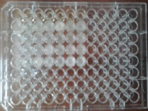
| Cells were seeded in 96-well plates at cell density 2 x 10⁵ cells/ well/ml. After 24 hours in media containing 10% FBS the media was changed to media containing the 10% FBS concentration [22,23]. |
| Cells were treated for 24 and 48 hours, and media was replaced with fresh media every 24 hour. Media was removed, and cells were washed with 200 μl/well PBS. 100 μl/well trypsin was added, and cells were incubated at 37°C for 10 minutes. The 96- well plates were tapped carefully to detach cells from the plate. Cells were harvested using 2 x 0.5 ml DMEM, and transferred to 1.5 ml eppendorf tubes for counting, fixing or freezing (Figures 4 and 5). |
|
MTT Assay
|
| 1. 100 μl of media was added into each of the 96 well plates from row B to row G (triplicate). |
| 2. Add 100 μl of diluted plant extract were added in row A and row B. |
| 3. Starting from row B , the 200 μl of solution (100 μl dilution + 100 μl media ) were mixed and 100 μl from row B were added into next row (row C ) by using micropipette and serial dilution was done upto row G |
| 4. Finally, excessive 100 μl from row G were discarded .The final volume for each well was 100 μl. |
| 5. Cultured MCF-7 cells were harvested by trypsinization, pooled in a 15 ml vial. |
| 6. The cells were plated at a density of 1×10⁶ cells/well (100 μl) into 96-well microtitre plates from row B to row G. |
| 7. Finally, 200 μl of cells were added in row H as a control. |
| 8. Each sample was replicated 3 times and the cells were incubated at 37°C in a humidified 5% CO2 incubator for 24 hours/48 hours. |
| 9. After incubation period, MTT (20 μl of 5 mg/ml) was added into each well and the cells incubated for another 2-4 hours until purple precipitates were clearly visible under microscope. |
| 10. The medium together with MTT (190 μl) were aspirted off the wells, DMSO (100 μl) was added and the plates shaken for 5 minutes. |
| 11. Absorbance for each well was measured at 540 nm. |
| By using, MTTS assay method for A-MCF 7 and similarly for B-MCF 7, different concentration where taken for different extracts likewise stock solution DMEM, concentration of dilutions and DMSO. Then we observed cytotoxicity effect of PSE and PPE on ELISA plate reader at 570 nm (ethanol), by reading the values we observed the PPE show’s better cancerous activity against the cancer then PSE as shown on graph of Figure 6. The graph demonstrates IC50 value in ethanol, no. of dillution v/s vaibility (Figure 6 and Tables 4-6) |
| The Results showed concentration-dependent reduction in the final number of cancer cells in consequence to treatment with the aforementioned plant extract. The anticancer effects were evaluated and contributed to the cytotoxic effects (decreased number of live cells). |
Conclusion
|
| The activity of Punica granatum was good against cancer cell line from this we concluded that PPE shows a better cancerous activity against the cancer as compare to PSE. |
Tables at a glance
|
|
|
Figures at a glance
|
 |
 |
 |
| Figure 1 |
Figure 2 |
Figure 3 |
 |
 |
 |
| Figure 4 |
Figure 5 |
Figure 6 |
|


