Research - (2023) Volume 17, Issue 1
External Protozoan Parasites of Sarowa Fish (Trachurus mediterraneus ) from Mediterranean Sea at Zliten Coastal Area, Libya
Samia Hamid Ahmed1* and
Radwan Eldehidya2
1Department of Fisheries and Wild life, College of Science and Technology of Animal Production, Sudan University of Science and Technology, Libya
2College of Marine Resources, Asmarya Islamic University, Zliten, Libya
Libya
*Correspondence:
Samia Hamid Ahmed, Department of Fisheries and Wild life, College of Science and Technology of Animal Production, Sudan University of Science and Technology,
Libya,
Email:
Received: 02-Jan-2023, Manuscript No. IPFS-22-13134;
Editor assigned: 04-Jan-2023, Pre QC No. IPFS-22-13134;
Reviewed: 18-Jan-2023, QC No. IPFS-22-13134;
Revised: 23-Jan-2023, Manuscript No. IPFS-22-13134;
Published:
30-Jan-2023, DOI: 10.36648/1307-234X.23.17. 101
Abstract
The aim of this study to conduct general survey of external protozoan parasites on (Sawrow) Trachurus mediterraneus from the Mediterranean Sea at coastal area of Zliten city, Lybia. Atotal of 12 specimens of fish species namely Trachurus mediterraneus were collected randomly. The fish were transported immediately alive to the laboratory in the Division of Marine Organisms and Fish Culture, College of Marine Resources, Asmarya University, where they were maintained alive in well aerated glass aquaria. External examination of each of fish for parasites was carried out. The study revealed different types of ciliates include Trichodina spp., Ichthyophthyrus spp., Epstylis spp., Chillodenella spp., Trichophyra spp. and Vorticella spp. and different type of flagellate which include, Amyloodinium spp., Cryptocaryon spp. Piscinoodinium spp. Vorticella spp.and Cryptobia spp. with density .83 in skin and .58 in the gill.
Keywords
Marine Protozoa; Fish; Costal Area; Zliten
INTRODUCTION
Aquaculture is currently one of the fastest growing food sectors
in the world, with the majority being finfish production. The total
world fish production is expected to reach 196 million tons (Mt)
by the year 2025, where aquaculture is estimated to surpass the
total production of capture fisheries. However, the capture sector
is expected to remain dominant for a number of fish species and
still be vital for supplying seafood both locally and globally (FAO,
2020). Fish is of importance in the diet of different countries
especially in the tropics and subtropics where malnutrition is a
major problem [1].
Wild-caught fish usually harbor several different parasites, as this
is a common occurrence in fish under natural conditions several
of these species show a high degree of host specificity and some
even have complicated life cycles involving different animal
species/types as intermediate hosts and are as such not directly
transmitted from fish-to-fish [2]. However, some being zoonotic
pathogens [3].
Parasitic infections often give an indication of the quality of water,
since parasites generally increase in abundance and diversity in more polluted waters. According to Klinger and Francis-Floyd
(2009) protozoa are a vast assemblage of eukaryotic organisms
and that most of the commonly encountered fish parasites
are protozoa. These parasites attack the fish, causing massive
destruction of skin and gill epithelium. Representatives within
the main protozoan groups such as amoebae, dinoflagellates,
kinetoplastid flagellates, diplomonadid flagellates, apicomplexans,
microsporidians and ciliates have been shown to cause severe
morbidity and mortality among farmed fish [4].
Trachurus mediterraneus are found usually near the bottom,
at times also in surface waters; pelagic and migratory in large
schools. They feed on other fishes especially sardines, anchovies
and small crustaceans. This is a commercial species. It can
be caught by various gears such as seines and fixed nets. The
contribution of Trachurus mediterraneus to local fisheries differs
in each sea. In the Black Sea, this species makes up 54% of the
total catch (2,919 t), whereas it makes up 39% in the Marmara Sea
(562 t), 4% in the Aegean Sea (247 t) and 3% in the northeastern
Mediterranean Sea.
The aim of this study to conduct general survey of external
protozoan parasites on (Sawrow) Trachurus mediterraneus from the Mediterranean Sea at coastal area of Zliten city, Lybia.
Materials and Method
Samples collection
Atotal of 12 specimens of fish species namely Trachurus
mediterraneus were collected randomly. Fishes were collected
by fishermen by using cast net and gill nets, during the period
from August 2016 until the end of May 2017 from Zliten coastal
area (Lat 32°30' N ; Long. 14°43' E) Modern harbour facility 156
km east of Tripoli; entrance shallow,.big.
The fish were transported immediately alive to the laboratory
in the Division of Marine Organisms and Fish Culture, College
of Marine Resources, Asmarya University, where they were
maintained alive in well aerated glass aquaria.
External examination
Visual examination of the body surface for external lesions,
ulcers, furuncles or granuloma especially fins, tail and gill for
proliferative and necrotic changes was done.
External examination of each of the fish for parasites was carried
out. The skin, gill and fins were also examined for ectoparasites by naked eye .Then wet smears were taken from the skin, fins and
gills and carefully were air dried, fixed in methanol for 10 min,
and then stained with 5% Giemsa’s solution in phosphate buffer
(pH 7.3) for 30 min. Smears were then examined using an Kruss
optronic microscope with an oil immersion lens.
Photography
Kruss optronic microscope with camera(BEL,Eurkm 10.0) was
used to photograph the parasite slide. The parasites were
identified using images from websites such and by making their
sketches as observed on the microscope and compared with the
pictorial guide on fish parasites by then identified.
Result and Discussion
The present study showed the existence of different stages of
species of external protozoans (Figures 1-7). The distribution
of the parasites, their location on the fish host body and mean
intensity of infection are summarized in Table 1.
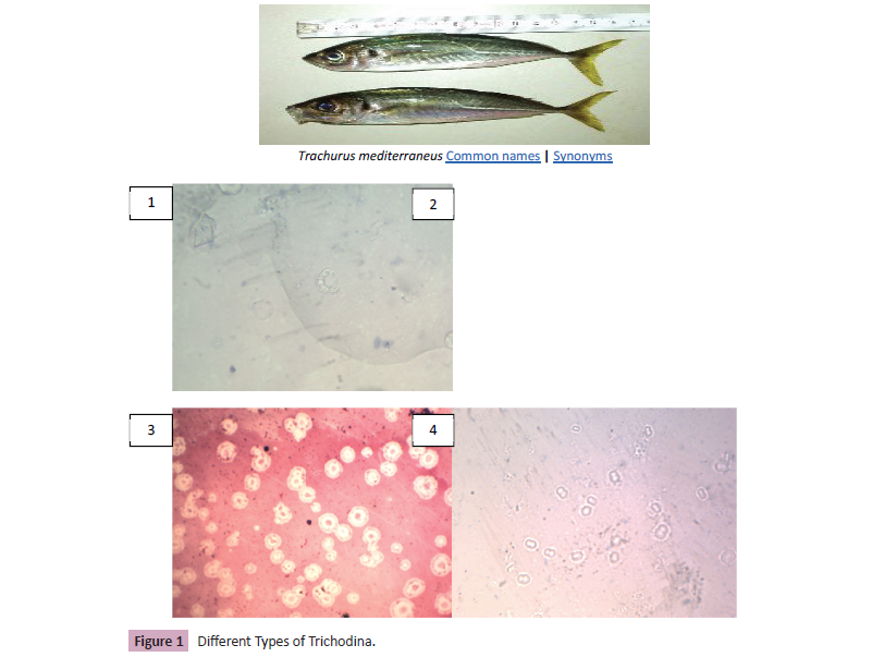
Figure 1: Different Types of Trichodina.
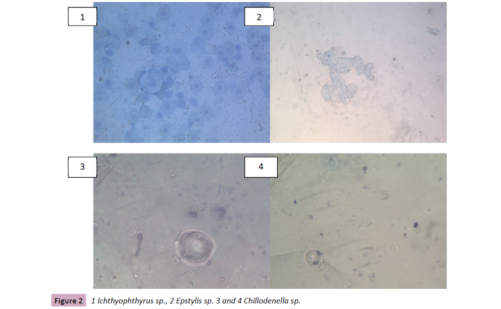
Figure 2: 1 Ichthyophthyrus sp., 2 Epstylis sp. 3 and 4 Chillodenella sp.
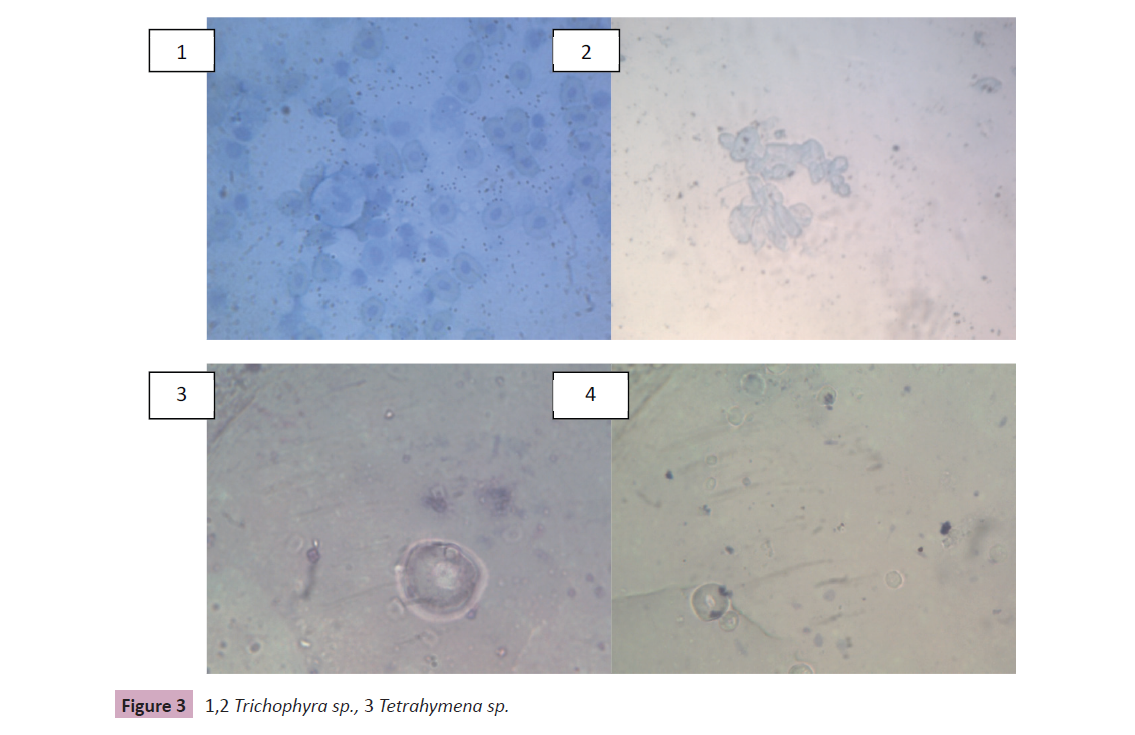
Figure 3: 1,2 Trichophyra sp., 3 Tetrahymena sp.
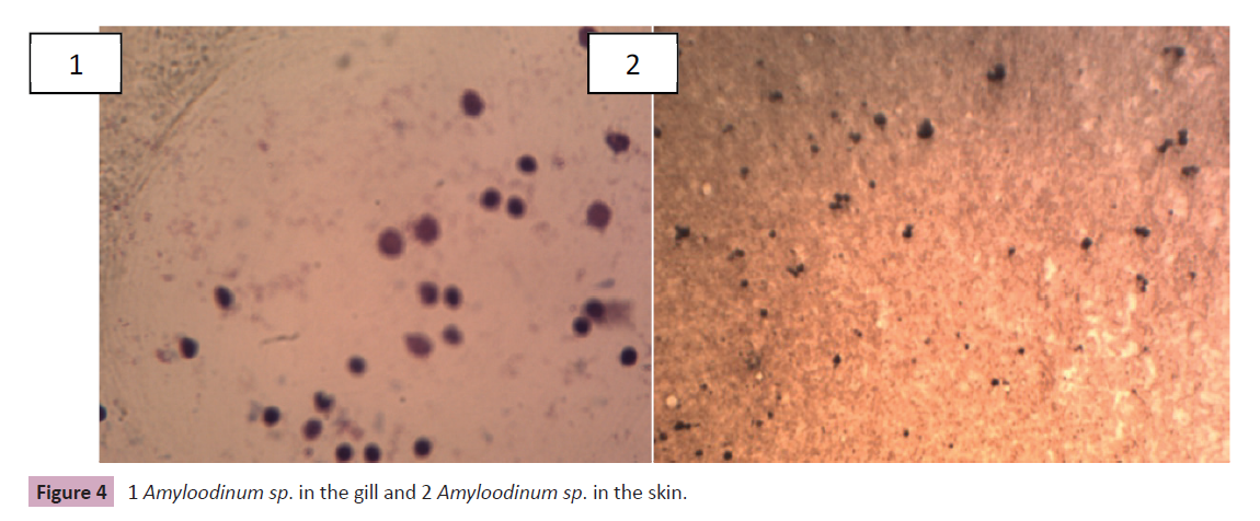
Figure 4: 1 Amyloodinum sp. in the gill and 2 Amyloodinum sp. in the skin.
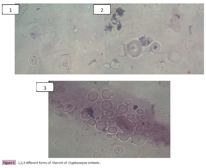
Figure 5: 1,2,3 different forms of theront of Cryptocaryon irritants .
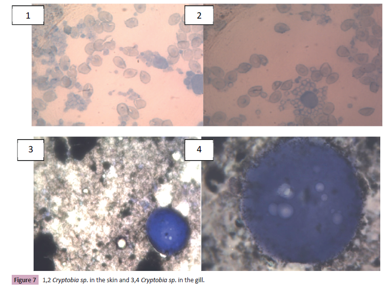
Figure 7: 1,2 Cryptobia sp. in the skin and 3,4 Cryptobia sp. in the gill.
| |
Site of infection |
| Parasites |
Skin |
gill |
| Trichodina spp. |
+ |
+ |
| Ichthyophthyrus spp. |
_ |
+ |
| Epstylis spp |
_ |
+ |
| Chillodenella spp. |
+ |
+ |
| Trichphyra spp. |
+ |
_ |
| Teteahymena spp. |
+ |
|
| Amyloodinium spp. |
+ |
+ |
| Cryptocaryon spp. |
+ |
_ |
| Piscinoodinium spp. |
_ |
+ |
| Vorticella spp. |
+ |
_ |
| Cryptobia spp. |
+ |
+ |
| Total |
10 |
7 |
| Density |
0.83 |
0.58 |
Table 1. List of parasites species found in the external organs.
According to [5] parasite of fish can either be external or internal.
Parasitic infections often give an indication of the quality of water,
since parasites generally increase in abundance and diversity in
more polluted waters and parasitic diseases are very common in fish all over the world and are of particular importance in the
tropics.
The study revealed different types of ciliates include Trichodina
spp., Ichthyophthyrus spp., Epstylis spp., Chillodenella spp.,
Trichophyra spp. and Vorticella spp. These parasites of worldwide distribution. This parasite is not host specific and fish can
potentially transmit the parasite. Most of the protozoan's
identified by aquaculturerists will be ciliates. Apiosoma,
Balantidium, Chilodonella, Epistyles, Ichthyophthirius multifiliis,
Tetrahymena and Trichodinidae are the main representatives [6].
Many of the parasites proliferate in organic debris accumulated in
the bottom of a tank or vat [7].
They use fish only for attachment and do not invade the epithelial
cells, thus feeding by filtration of suspended material in the
water. This phenomenon is termed epibiosis, in which the ciliate
acts as epibiont and the host as basibiont (substrate organism)
[8]. When present in a population under high stocking densities
associated with bacteria that colonizes its peduncle, this ciliate
might cause host damage and then be named as parasite with
greater impact in farmed catfish [9].
These include the following, Trichodinids can be found parasitizing
both freshwater and marine fishes on the body surface, buccal
cavity and gills. Nevertheless, relatively few of them have become
endoparasites in the intestine, kidney and urinary bladder of
their hosts (Lom and Dykova, 1992). Differently, some trichodinid
species were found to be suppressed with increased water
temperature.
Heavy infestations of juvenile lumpfish with Trichodina sp. have
been reported. The parasites are a commonly reported finding
in Norwegian aquaculture, but are not usually related to major
health problems. Trichodinids have also been found in varying
abundance on the gills of wild-caught wrasse, usually without
any associated pathology. In a short period of time the increased
number of parasites per host as observed.
Ichthyophthyrus spp. which causes disease called "Ich" or "white
spot disease" has been a problem to aquarists for generations.
The causative agent of ichthyophthiriasis or white spot disease
is one of the most important fish parasites of worldwide
distribution compromising skin, fins, gills and eyes of farmed
fish. This parasite is not host specific and any freshwater fish can
potentially transmit the parasite.
Epistyles are a stalked ciliate that attaches to the skin or fins of the
host. Epistyles are of greater concern than many of the ciliates
because it is believed to secrete proteolytic ("protein-eating")
enzymes that create a wound, suitable for bacterial invasion, at
the attachment site. Also Chilodonella species are free-living but
some of them parasitize the skin, gills and fins of both freshwater,
marine and estuary fish.
Other ciliates have been reported from the gills of wrasse without
any apparent damage to the tissue, such as the Cryptocaryon-like
ciliates. The species C. irritans is known as the cause of ‘white
spot’ disease in marine fish. Similarly, protozoan ciliates have
been found on Atlantic salmon.
According to, these ciliates do not form cysts and studies suggest
that Tetrahymena penetrates the host epithelium (especially
where there are wounds) reaching the blood and parasitizing the
gills, kidney, eyes and brain. High organic loads and deterioration
of water quality are often associated with heavy, debilitating
Ambiphrya infestations.
Under a microscope, it can be observed three forms of the dinoflagellate from the scraps of fish: pear-shaped, bananashaped
and the mature rounded parasite of brownish color.
although in high infestations different developmental stages can
be found. Special care must be taken on the mature trophonts
that could be confused with I. multifiliis by an inexperienced
person.
Amyloodinium comprises dinoflagellates of varied shape
depending on the life stage. The causative agent of velvet disease
A. ocellatum is ubiquitous, affects marine farmed fish and may
provoke important outbreak mortalities and economical losses in
aquaculture systems.
Piscinoodinium is a sedentary flagellate that attaches to the
skin, fin, and gills of fish. The common name for Piscinoodinium
infection is "Gold Dust" or "Velvet" Disease. The parasite has an
amber pigment, visible on heavily infected fish. Affected fish will
flash, go off feed, and die. Piscinoodinium is most pathogenic to
young fish.
Cryptobia is a flagellated protozoan common in cichlids Cryptobia
typically is associated with granulomas in which the fish "walls
off" the parasite [10]. These parasites have been observed
primarily in the stomach, but may be present in other organs.
Some organisms are commensals rather than true parasites. For
example, Cryptobia dahli is a flagellate commonly found within
the stomach of wild-caught lumpfish. It is regarded as harmless
for the lumpfish, since it does not cause any apparent damage to
the gastric tissue, even when present in extremely high numbers.
As such, it is not considered to become a problem for lumpfish
aquaculture in the future.
Parasitism is a key component in all ecosystems, playing a
fundamental role at the population level and wider ecological
scales. Although parasites play a key role in food web interactions,
their diversity, dynamics, and influence on ecosystems remain
neglected.
References
- Bruno DW,Nowak B,Elliott DG(2006)Guide to the identification of fish protozoan and metazoan parasites in stained tissue sections.Diseases of Aquatic Organisms70:1-36.
Indexed at, Google Scholar, Crossref
- Buchmann K(2015) Impact and control of protozoan parasites in maricultured fish. Parasites in Fisheries and Mariculture (124): 168-177.
Indexed at, Google Scholar, Crossref
- https://www.researchgate.net/publication/264789298_Infectious_diseases_of_warmwater_fish_in_marine_and_brackish_waters
- Dyková I,Tyml T,Kostka M,Pecková H(2010)Strains ofUronema marinum(Scuticociliatia) co-isolated with amoebae of the genus Neoparamoeba.Diseases of Aquatic Organisms89:71-77.
Indexed at, Google Scholar, Crossref
- Eiras JC, Takemoto RM, Pavanelli GC, Luque JL (2012) Checklist of protozoan parasites of fishes from Brazil. Zootaxa 3221: 1-25.
Google Scholar
- https://io.furg.br/images/PARASITOS-PEIXES-MARINHOS-AMERICA-SUL-EBOOK.pdf
- https://www.fao.org/3/ca9229en/ca9229en.pdf
- Freeman MA,Kristmundsson A(2018) A closer look atCryptobia dahli: a parabodonid flagellate from the stomach of the Atlantic lumpfish.Bulletin of the European Association of Fish Pathologists38:195-201.
Google Scholar
- https://www.vetinst.no/rapporter-og-publikasjoner/rapporter/2019/fiskehelserapporten-2018
- Ishikawa MM (2012) Trichodina heterodentata(Ciliophora: Trichodinidae): a new parasite for (Pisces: Characidae).Piaractus mesopotamicus, Zootaxa3422(2012): 62-68.
Google Scholar
Citation: Ahmed SH, Eldehidya R, Elaakary S (2023) External Protozoan Parasites of Sarowa Fish (Trachurus mediterraneus ) from Mediterranean Sea at Zliten Coastal Area, Libya. J Fish Sci, Vol.17 No. 1: 101.
