Introduction
All surgical procedures carry a risk of forgetting any foreign material within a cavity; the increase of the population has conditioned saturation of the health sector, performing about 16 surgeries per surgeon per month, 30% of these are emergency surgeries, giving the surgeon a high probability of this complication [1]. Textiloma can be defined as anymaterial used during surgery that is forgotten in the body, it is also known as as gossypiboma, a word derived from the Latin Gossypium (cotton) and kiswahiliboma (confinement);are a group of pseudotumor caused by non-absorbable surgical material without any therapeutic effect [2,3]. Generate many of the times, an increase in the days of hospital stay and increase costs of health care and serious complications in surgical patients, as well as legal problems. The actual incidence of this complication is unknown, reports of this cases is done sporadically, worldwide with an incidence of 1 in 1500-3000 surgeries performed, with an average age of 20-50 years, highly related to surgeries that involve abdominal and thoracic cavity (52%), although cases have also been reported in gynecological surgery (22%), urology (10%), vascular (10%), orthopedic (6%) and neurosurgery (6%) [4- 6]. The diagnosis is based on the patient's surgical history with localized pain and auxiliary medical imaginging which the foreign material can be shown. The treatment is based on the removal of the same and the resolution of the complications associated with their presence [7-9]. This text present the case of 40-yearold femalewith multiple surgical history with abdominal pain since one monthlocated in the right iliac fossa, exacerbated nine days before admission, showing in imaging studies textiloma (compress) which is removed laparoscopically in its entirety.
Case Report
A 40-year-old female patient with a surgical history of congenital hip dislocation which required 13 reconstructive surgeries, the last with complete replacement of the right hip, laparoscopic appendectomy 1 year ago, C-section and bilateral tubal occlusion six months ago, who enters to emergency department for abdominal pain in both lower quadrants of a month of evolution associated with distended abdomen, and nausea without vomiting, treated with conventional analgesics without clinical improvement, exacerbated nine days before admissionwith increasing pain becoming a 10/10 intensity referred by family doctor, with a palpable mass in the right iliac fossa. During the physical examination, wiht facies of pain and antalgic position, vital signs with a tendency to tachycardia and fever, distended abdomen with pain on deep palpation in both lower quadrants, no evidence of peritoneal irritation, palpable mass 8 by 8 cm of solid consistency, mobile, decreased peristalsis.
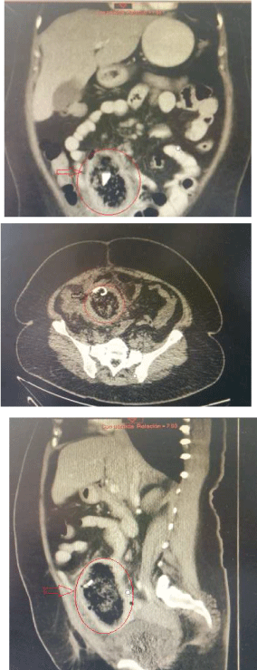
Laboratories: leukocytes 9.18 xmm3, hemoglobin 11.2 g/dl, hematocrit 35.3%, 419 platelets, creatinine 0.7 mg/dl, 82 mg glucose/dl , sodium 138 mEq/L, potasisium 4.3 mEq/L, 107 chlorine mEql/L; abdominal radiography without evidence of intestinal perforation, distended loops and air-fluid levels in the iliac fossa, CT requested for suspected tumor dependent of the cecum, with right lower quadrant image with linear hyperdense material, peripheral inflammatory reaction,suggestive of textiloma (Figure 1).

Figure 1: Right lower quadrant image with linear hyper dense material, peripheral inflammatory reaction, suggestive of textiloma.
Accessing to the service of Surgery with antimicrobial impregnation and programming surgery, wich is performed laparoscopically with a 12mm trocar supraumbilical and 2 side of 5 mm and 10 mm respectively, in the diagnostic laparoscopy found multiple adhesions of the omental to wall which are released finding a mass of intestinal loops and omentum with abundant inflammatory tissue in cecum region, performing the dissection with instrument obtaining 800 ml of pus, which is sucked observing in the bottom the material, corresponding to a surgical compress which It is released from its attachments by making hydrodissection removing entirely by port of 12 mm, intestinal loops which are presented without macroscopic evidence of intestinal perforation, placing a directly Penrose drain into the mass.
Three days after that procedure is presented with abdominal pain, fever and leukocytosis, for this is taked to surgery for exploratory laparotomy, finding bowel perforation to 1.40 m of the angle of Treitz with necrotic edges and eversion of the mucosa, so they decide to make jejunostomy and washing cavity with good clinical outcome after surgery and discharged home with stoma scheduled for restoration of intestinal transit event.
Discussion
Textiloma is a foreign surgery textile material within a cavity duriing the surgical event, it has a relatively frequent occurrence in the emergency service and represents a differential diagnosis in all patients with surgical history and abdominal pain. It has the highest incidence in the population of 20-50 years and are directly related to emergency surgeries, globally there is no specific frequency but is accepted that it occurs in one in 1500 to 3 000 abdominal surgeries.
Approximately 80% of the foreign bodies correspond to compresses; bandages and surgical drapes (“gossypibomas”) containing indigestible cellulose fibers by the human body. Among the known risk factors are emergency surgeries, the surgeries that are done in night shift, abrupt changes in plan sor surgical technique, lack of leadership surgeon, cell phone use during the surgical event, major bleeding, patient with high body mass index.
The order of frequency in surgeries involving the abdominal and thoracic cavity (56%), although cases have been reported in gynecological surgery (22%), urology (10%), vascular (10%), orthopedic (6%), and neurosurgery (6%).
The sequence of events that is presented after 24 hours is exudative inflammation; the eighth to thirtheenth day exists granulomatous inflammation that is a distinctive pattern of chronic inflammation characterized by aggregation of actived macrophages, these macrophages acquire an aspect squamous cells enlarged surrounded by lymphocytes, fibroblasts and connective tissue, who`s function is to contain the offending agent; these cells cause adhesión to the surrounding tissues. After 5 years, they may eventually desintegrate, calcify and, less frequently, ossify.
Due to the gossypibomas material that is non-absorbable and does not exist any descompantation in the body, and generates 2 different types of reactions: the first kind is a response aseptic fibronosa that generates adherence and conditions an encapsulate with the formation of a foreign body granuloma. The presented symptoms are generated in patients with a reaction to intestinal oclusión [10-12].
The second reaction is of type exudative that will form an abscess with or without bacterial infection. In additional this may also cause to the formation of a fistula that will drain the content to hollow viscera or to the exterior [13]. The symptomatology can vary since abdominal pain, fever, intestinal obstruction, palpable pseudotumor, bleeding of the digestive tube, episodes of acute abdomen; some may even appear asymptomatic found accidently in routine laboratory studies.
Some of its possible complications are secondary sepsis due to the inflammatory process, localized abscesses, migration of textile material due to transmural erosion, peripheral adherence up to 68% of the cases, like infertility and vascular lesions. A 22% has been reported with intestinal perforation just like the formation of fistulas from the intestine to the skin [14].
The diagnosis is based on clinical history up to 80%, can be diagnosed by a simple abdominal x-ray, since most textile materials is radiopaque, however the process of phagocytosis and enzymatic digestion as well as depth and time evolution may disintegrate these materials and not be visible until overlooked. Ultrasound is an imaging study that can project a peripheral cystic mass with posterior acoustic shadowing and hypoechoic margins as well as peripheral liquid or intra-abdominal fluid collections. The Computed Axial Tomography evidence in acute gossypibomas a heterogeneous mass containing large amount of trapped air, which may or may not be surrounded by a peripheral blade ring, while chronic gossypibomas resemble a tumor that does not capture the intravenous contrast, with calcifications simulating a solid mass with air entrapped gas bubbles so that persist over time, this finding to the Computed Axial Tomography is considered the most specific for the detection of Gossypiboma [15,16].
It may be displayed in studies such as magnetic resonance and endoscopy as well as intestinal transit filling defects [17-20]. Treatment is based on surgical removal of the gossypiboma and resolution of associated complications. Open surgery is preferred by most surgeons because it generates adequate exposure and direct view of the anatomical site, however due to the advent of laparoscopic surgery many of these gossypibomas can be removed by this technique, which offers the advantage of magnification of the display as well as early recovery and declining associated morbidity involved in open surgery.
There are multiple reports of laparoscopic extraction of gossypibomass or diagnostic laparoscopy with conversion to open surgery most of them gauze or residual textile material, the present case reports presents one of the first reported cases of a compress fully extracted by laparoscopy. It is reoperated in a second surgical time by intestinal perforation which was not evident in the first intervention.
Conclusions
The knowledge of this pathology and its presence as a differential diagnosis is essential in the handling of surgical patients. Careful surgical technique and respect for international standards control of the surgical patient have a direct impact on the reduction of this type of complication. It is proposed that the laparoscopic procedure as the diagnostic method of direct vision as well as resolution method if gossypiboma is in stable patients without evidence of complications in centers where there is an expert laparoscopic surgeon. The lower morbidity and mortality of laparoscopic versus open procedures keep evidence for use as a first step in the treatment of gossypibomas.
8483
References
- Hyslop JW, Maull KI (1982) Natural history of the retained surgical sponge. South Med J 75: 657-660.
- Gawande A, Studdert D, Orav J, Brennan TA, Atul A, et al. (2003) Risk Factors for Retained Instruments and Sponge safter Surgery. N Engl J Med 348: 229-235.
- Asuquo ME, Ogbu N, Udosen J, Ekpo R, Agbor C, et al. (2006) Acute abdomen from gossypiboma: A case series and review of literature.Nigerian Journal of Surgical Research 8: 174-176.
- Kiernan F, Joyce M, Byrnes CK, O´Grady H, Keane FB, Nearyp (2008) Gossypiboma: a case report and review of the literature. Ir J Med Sci 177: 389-391.
- Custovic R, Krolo I, Marotti M, Babie N, Karapanda N (2004) Retained Surgical Textilomas occur more often during war. Croat Med J 45: 422
- Noriyuki Y, Kiyokazu N (2008) Intra-abdominal textiloma. Intra-Abdominal Textiloma. A retained surgical sponge mimicking a gastric gastrointestinal stromal tumor: report of a case. Surg today 38: 552–554.
- Taçyildiz I, Aldemir M (2003) The mistakes of surgeons: “gossypiboma”. Acta Chir Belg 103: 71-75.
- Haegeman S, Maleux G, Heye S, Daenens K (2008) Textiloma complicated by abscess-formation, three years after surgical repair of abdominal aortic aneurysm. JBR-BTR;91: 51-53.
- Dhillon JS, Park A (2002) Transmural migration of a retained laparotomy sponge. Am Surg 68: 603-605.
- El Khoury M, Mignon F, Tarvidon A, Mesuralle B, Roachard F, et al. (2002) Retained surgical sponge gossypiboma of the breast. Eur J Radiol ;42: 58-
- Robbins S, Kumar V, Cotran R (1999) Patología humana. 6ª edición. México (D.F.): Mc Graw-Hill Interamericana Editores; p.46-47.
- Esposito S, Ragozzino A, Rossi G, Pinto A, Martino A (1994) Spontaneous migration of a surgical sponge in the small intestine. Apropos of a case studied with conventional radiology and CT. Radiol Med (Torino) 88: 139.
- Sugano S, Suzuki T, Iimura M, Mizugami H, Kagesawa M, et al. (1993) Gossypiboma: diagnosis with ultrasonography. J Clin Ultrasound 21: 289-292.
- Cheng TC, Chou AS, Jeng CM (2007) Computed Tomography Findings of Gossypiboma. J Chin Med Assoc 70: 565-569.
- Gencosmanoglu R, Inceoglu R (2003) An unusual cause of small bowel obstruction: gossypiboma - case report. BMC Surgery 3: 1-6
- Cerwenka H, Bacher H, Kornprat P, Mischinger HJ (2005) Gossypiboma of the liver: CT, MRI and intraoperative ultrasonography findings. Dig Surg 22: 311-312.
- Kokubo T, Itai Y, Ohtomo K, Yoshikawa K, Lio M, et al. (1987) Retained surgical sponges: CT and US appearance. Radiology 165: 415-418.
- Vega GR, Heredia NM, Camacho P, Tenorio M, Barreda J, et al. (2002) Extracción de un cuerpo extraño por cirugía laparoscópica. Reporte de un caso y revisión de la literatura. Revista Mexicana de Cirugía Endoscópica 3: 175.


