Image - (2015) Volume 3, Issue 2
Glucocorticoid Injection-Induced Cutaneous Atrophy
Paloma Vela Casasempere*
Rheumatology Department, Hospital General Universitario de Alicante, Universidad Miguel Hernández, Maestro Alonso 109, Alicante, Spain 03010
*Corresponding Author:
Paloma Vela Casasempere
Rheumatology Department
Hospital General Universitario de Alicante, Universidad Miguel Hernández
Tel: +34 965933015
Fax: +34 965913786
E-mail: palomavela62@gmail.com
Abstract
De Quervain’s tenosynovitis consists on inflammation and stenosis of the tendon sheath enveloping the abductor pollicis longus and the extensor pollicis brevis as they course through the sheath at the level of the radial styloid process. Glucocorticoid injection is commonly used to treat this condition, besides other soft-tissue rheumatic disorders.
Clinical Image
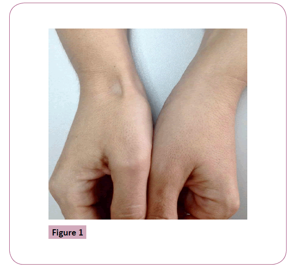
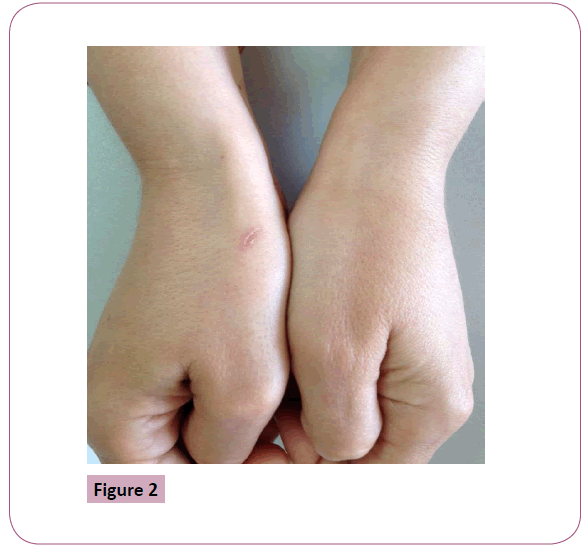
De Quervain’s tenosynovitis consists on inflammation and stenosis of the tendon sheath enveloping the abductor pollicis longus and the extensor pollicis brevis as they course through the sheath at the level of the radial styloid process. Glucocorticoid injection is commonly used to treat this condition, besides other soft-tissue rheumatic disorders. This technique offers the advantage of high persistent local concentrations of corticoid in the desired area without significant systemic absorption and serious side effects. Cutaneous atrophy is among the most frequent adverse effect following injected corticosteroid, usually begins within 2 to 3 months of injection and resolves spontaneously within 1 to 2 years [1]. The exact mechanism that makes it appear is not fully understood, but is likely due to several factors, as decreased synthesis of type I collagen, degeneration of sebaceous glands, decreased elastin, epidermal atrophy, and involution of subcutaneous fat lobules [2]. This patient developed a local cutaneous atrophy two months after glucocorticoid injection to treat De Quervain’s tenosynovitis (Figure 1), which was spontaneously resolved six months later (Figure 2).

Figure 1

Figure 2
6493
References
- Dyment PG (1970) Local atrophy following triamcinolone injection. Pediatrics 46: 136-137.
- Fülöp E (1976) Mechanism of local skin atrophy caused by intradermally injected corticosteroids. Dermatologica 152: 139-146.


