Lvming Zhang1, Zhao Dong2,Huidan Fu3 and Shengyuan Yu2*
Department of Neurology, Aerospace Center Hospital/Aerospace Clinical Medical College Affiliated to Peking University, Beijing, China
Department of Neurology, Chinese PLA General Hospital, Beijing, China
Department of Neurology, The New district People’s Hospital of Luoyang, Henan Province, China
*Corresponding Author:
ShengYuan Yu
Department of Neurology
Chinese PLA General Hospital
Fuxing Road 28, Haidian District
Beijing 100853
China
Tel: +86 10 6688 7329
E-mail: yusy1963@126.com
A 52-year-old man with a history of diabetes mellitus presented with acute horizontal diplopia and transient vertigo for 6 days. Neurologic examination revealed isolated left abducent limitation and right positive Babinski sign with no limbs weakness. The rest of his extraocular movements were normal, and no nystagmus or internuclear ophthalmoplegia was observed. He did not have additional pontomedullary symptoms or signs, such as facial palsy, sensory loss, dysarthria or weakness.
A 52-year-old man with a history of diabetes mellitus presented with acute horizontal diplopia and transient vertigo for 6 days. Neurologic examination revealed isolated left abducent limitation (Figure 1A) and right positive Babinski sign with no limbs weakness. The rest of his extraocular movements were normal, and no nystagmus or internuclear ophthalmoplegia was observed. He did not have additional pontomedullary symptoms or signs, such as facial palsy (Figure 1B), sensory loss, dysarthria or weakness. A brain diffusion-weighted MRI scan showed a hyperintense signal in the left ventral pons (Figure 1C) in the localized region of the abducent nerve fascicle. Our case showed that localized pontine infarction may mimic an isolated abducent nerve palsy which had been rarely reported (Figure 1D) [1,2].
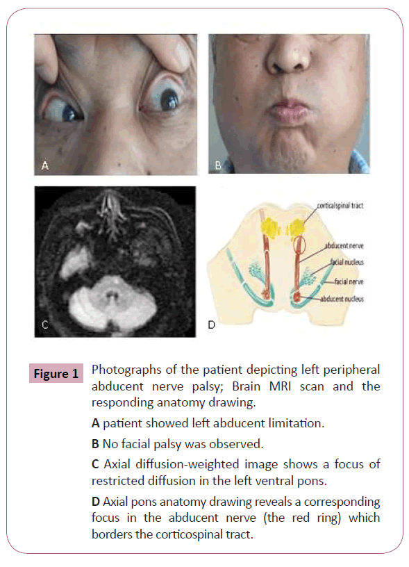
Figure 1: Photographs of the patient depicting left peripheral abducent nerve palsy; Brain MRI scan and the responding anatomy drawing.
A patient showed left abducent limitation.
B No facial palsy was observed.
C Axial diffusion-weighted image shows a focus of restricted diffusion in the left ventral pons.
D Axial pons anatomy drawing reveals a corresponding focus in the abducent nerve (the red ring) which borders the corticospinal tract.
7300
References
- Jong WP, Suk YK, Young HS (2004) Isolated Abducens Nerve Palsy due to Anterolateral Pontine Infarction. Eur Neurol 52: 254–256.
- Toshio Fukutake, Keizo Hirayama (1992) Isolated abducens nerve palsy from pontine infarction in a diabetic patient. Neurology 11: 2226.


