Clinical Images - (2017) Volume 5, Issue 2
Liposarcoma of the Pelvis and Soft Tissues
Sarah El-Abbassi*, Hasnae Sfaoua, Tayeb Kebdani and Noureddine Benjaafar
Department of Radiation Oncology, National Institute of Oncology, Mohammed V University in Rabat, Morocco
*Corresponding Author:
Sarah El-Abbassi
Resident, Department of Radiation Oncology
National Institute of Oncology
Mohamed V University in Rabat, Morocco
Tel: +212661678389
E-mail: drsarahelabbassi@gmail.com
Received Date: April 26, 2016; Accepted Date: April 28, 2017; Published Date: April 30, 2017
Citation: El-Abbassi S, Sfaoua H, Kebdani T, et al. Liposarcoma of the Pelvis and Soft Tissues. Arch Can Res. 2017, 5:2. DOI: 10.21767/2254-6081.1000i31
Abstract
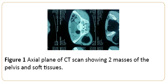
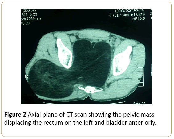
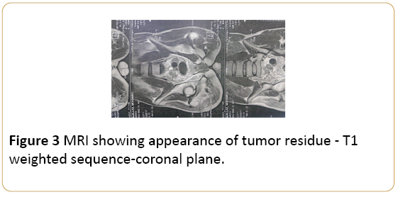
A 49-year-old man presented in the hospital with a large mass of the right buttock slowly growing for 2 years. It was associated to worsening low back pain radiating to the posterior aspect of the right hip, buttock and down to the postero-lateral aspect of the left foot. It was aggraved by sitting and walking. The patient presented also chronic constipation, dysuria and pelvic pain. A loss of appetite with weight loss was noted too. On physical assessment, a large tumor deforming the right buttock was found. It was measuring 20 cm long axis. Pelvis palpation showed a painless mass measuring 10 cm long axis simulating a full bladder. The pelvic CT scan showed a large mass of the right buttock measuring 250 mm × 140 mm. It was extending inside the pelvis through the sciatic notch. It was also well limited. The pelvic component was measuring 120 mm × 106 mm, deplacing the rectum to the left, bladder anterioly. The CT density of the mass was that of fat. Except at the posterior aspect, where there was an irregular area of higher CT density. In the view of higher CT density in one area, the diagnosis of liposarcoma was strongly suspected. No bone lysis or lymphadenopathy was found (Figures 1 and 2). CT scan of the chest and abdomen was negative for distal metastasis. The case was discussed in multidisciplinary oncological team and primary surgery was planned. At surgery, a large pelvic mass herniating through the right greater sciatic notch into the left buttock and thigh was found. Patient underwent a surgical resection. The postoperative histopathological findings confirmed the diagnosis of liposarcoma lipoma like with positive surgical margins. One month after surgery, a Pelvic Magnetic Resonance Imaging (MRI) was performed. It showed a tumor residue measuring 36 mm × 28 mm (Figure 3). No surgical recovery was done. External beam radiotherapy of tumor bed at a radiation dose of 2 Grays (Gy) /fraction given five times weekly (Monday to Friday) to a dose of 50 Gy. A boost at the dose of 15 Gy was delivered to the tumor residue. Patient completed the treatment without any major untoward event. At the time this report was written, the patient had 6 months of follow-up. No evidence of malignacy is found.
Clinical Image
A 49-year-old man presented in the hospital with a large mass of the right buttock slowly growing for 2 years. It was associated to worsening low back pain radiating to the posterior aspect of the right hip, buttock and down to the postero-lateral aspect of the left foot. It was aggraved by sitting and walking. The patient presented also chronic constipation, dysuria and pelvic pain. A loss of appetite with weight loss was noted too. On physical assessment, a large tumor deforming the right buttock was found. It was measuring 20 cm long axis. Pelvis palpation showed a painless mass measuring 10 cm long axis simulating a full bladder. The pelvic CT scan showed a large mass of the right buttock measuring 250 mm × 140 mm. It was extending inside the pelvis through the sciatic notch. It was also well limited. The pelvic component was measuring 120 mm × 106 mm, deplacing the rectum to the left, bladder anterioly. The CT density of the mass was that of fat. Except at the posterior aspect, where there was an irregular area of higher CT density. In the view of higher CT density in one area, the diagnosis of liposarcoma was strongly suspected. No bone lysis or lymphadenopathy was found (Figures 1 and 2). CT scan of the chest and abdomen was negative for distal metastasis. The case was discussed in multidisciplinary oncological team and primary surgery was planned. At surgery, a large pelvic mass herniating through the right greater sciatic notch into the left buttock and thigh was found. Patient underwent a surgical resection. The postoperative histopathological findings confirmed the diagnosis of liposarcoma lipoma like with positive surgical margins. One month after surgery, a Pelvic Magnetic Resonance Imaging (MRI) was performed. It showed a tumor residue measuring 36 mm × 28 mm (Figure 3). No surgical recovery was done. External beam radiotherapy of tumor bed at a radiation dose of 2 Grays (Gy) /fraction given five times weekly (Monday to Friday) to a dose of 50 Gy. A boost at the dose of 15 Gy was delivered to the tumor residue. Patient completed the treatment without any major untoward event. At the time this report was written, the patient had 6 months of follow-up. No evidence of malignacy is found.

Figure 1: Axial plane of CT scan showing 2 masses of the pelvis and soft tissues.

Figure 2: Axial plane of CT scan showing the pelvic mass displacing the rectum on the left and bladder anteriorly.

Figure 3: MRI showing appearance of tumor residue - T1 weighted sequence-coronal plane.
19142


