Wilson Onuigbo1* and Obioma Okezie2
1Department of Histopathology, University of Nigeria Teaching Hospital, Nigeria
2Department of Obstetrics and Gynaecology, University of Nigeria Teaching Hospital, Nigeria
Corresponding Author:
Wilson Onuigbo
Department of Histopathology, University of Nigeria Teaching Hospital, Enugu 400001, Nigeria
Tel: 2348037208680
E-mail: wilson.onuigbo@gmail.com
Received date: September 20, 2016; Accepted date: October 26, 2016; Published date: October 31, 2016
Citation: Onuigbo W, Okezie O. Liposarcoma of the Uterine Cervix in Nigeria: Case Report. Arch Can Res. 2016, 4: 4. doi: 10.21767/2254-6081.1000110
Keywords
Uterus; Cervix; Liposarcoma; Epidemiology; Nigeria
Introduction
A massive review of uncommon sarcomas of the uterine service was carried out by Fadare [1]. Concerning liposarcoma, it was concluded that they are “extraordinarily rare.” Moreover, it was stated that “only four cases of pure liposarcoma of the cervix have been reported in the English literature.” These four had an average of 54 years (range 45– 62). Therefore, it is of interest that the local case about to be presented was aged 65 years. Its appreciation was facilitated by a Birmingham (UK) group [2], which postulated that a histopathology data pool can facilitate the prosecution of epidemiological analysis. Such a pool, established by the senior author (WO), was used here and is deemed to be worthy of documentation.
Case Report
NI, a 65-year-old, Para 8, Nigerian woman of the Igbo Ethnic Group [3], consulted one of us (OO) with the complaint of bleeding through the vagina for 3 months. She had been postmenopausal for 10 years and the cervix was tumorous. After obtaining her consent, the necessary preliminary investigations were carried out. As all were negative, cervical cone biopsy was carried out.
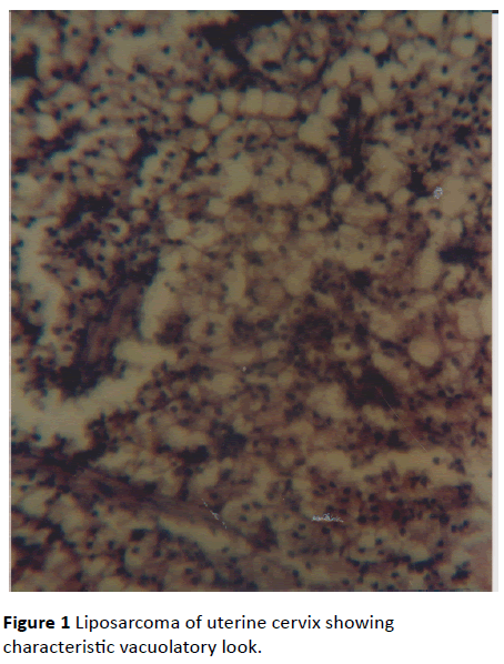
Numerous crumbly pale masses up to 3.5 cm across were obtained and submitted to the Senior author (WO). At microscopy, after routine processing, normal tissue was not seen. Rather, there was replaced by a sarcomatous growth whose vacuolatory differentiation was striking. See accompanying Figure 1. Liposarcoma was therefore diagnosed. Unfortunately, as so often happens in the locality [4], she was lost to follow-up.

Figure 1 Liposarcoma of uterine cervix showing characteristic vacuolatory look.
Discussion
Literature search revealed some recent single case reports including the one from the Northern Region of this country, Nigeria [5]. Aged 45 years, her lipomatous growth was fungating and was deemed to be probably the first documented in the African literature.
Elsewhere, Karateke’s group [6] reported the case of the 23- year-old woman. She was presented as the first case detected as having occurred in the productive period.
From Japan, Takeuchi et al. [7] described the 49-year-old woman whose case was distinguished on the ground that “Most of the adipocytes and vacuolated lipoblasts were positive for S-100 protein.” Perhaps, I should add here the group [8] that considered their patient as being “the exceptionally rare case of a low-malignant liposarcoma of the uterus.”
In the case from Poland [9], the woman was aged 71 years and had isolated liposarcoma of the uterine corpus in association with preinvasive cervix cancer. From India [10], liposarcoma was regarded as the least common of the uterine cervical sarcomas, the others being, in descending order, embryonal rhabdomyosarcoma, leiomyosarcoma, undifferentiated endocervical sarcoma, alveolar soft part sarcoma, and Ewing’s sarcoma.
The only case with clear antecedent was that in which Tamoxifen treatment was mentioned [11]. In our case, such esoteric treatment did not take place. Unfortunately, follow up was not possible.
17328
References
- Fadare O (2006) Uncommon sarcomas of the uterine cervix: a review of selected entities. Diagnost Pathol 1:30.
- Macartney JC, Rollaston TP, Codling BW (1980) Use of a histopathology data pool for epidemiological analysis. J Clin Pathol 33: 351-353.
- Kotwal PP, Faruoque M (1998) Macrodactyly. J Bone Joint Surg 80: 651-653.
- Obafunwa JO, Uguru VE (1999) Liposarcoma of the cervix. Trop Geog Med 42: 90-91.
- Karateke A, Gurbuz A, Kabaca C (2005) Uterine liposarcoma in a young woman: a case report. Int J Gynecol Cancer 15: 1230-1234.
- Takeuchi K, Murata K, Funaki K (2000) Liposarcoma of the uterine cervix: case report. Euro J Gynaecol Oncol 21: 290-291.
- Schneebauer J, Brinninger G, Halabi M, Nagl F (1996) Liposarcoma of the uterus. Gynakologisch-geburtshilfliche Rundschau 36: 90-91.
- So?nik H, Jele? M, So?nik K, Pomorska M (2006) Liposarcma of the uterine corpus coexisting with preinvasive cervical cancer–a case report. Pol J Pathol 57: 171-173.
- Khosla D, Patel FD, Kumar R (2013) Sarcomas of the uterine cervix: A united and multidisciplinary approach is required. Womens Hlth 9: 501-504.
- Hummel LP, Xiao-Jun W, Hean-Pierre G (2003) Pleomorphic liposarcoma of the uterus: Case report and literature review. Int J Gynecol Pathol 22: 407-411.


