Case Blog - (2015) Volume 3, Issue 2
Mohamad Khawandanah MD* and Mohamad Cherry MD Hematology-Oncology Section, Department of Medicine, The University of Oklahoma Health Sciences Center, Oklahoma City, OK, USA |
Corresponding Author: Mohamad Khawandanah MD, Assistant Professor of Medicine, University of Oklahoma Health Sciences Center Stephenson Cancer Center, 800 NE 10th street, Oklahoma City, OK 73102, USA, Tel: 1405-271-4000 E-mail: Mohamad-khawandanah@ouhsc.edu |
Related article at Pubmed, Scholar Google |
44-year-old female patient presented to our facility with 2 week history of headaches and massive mass located in the right forehead. She was diagnosed with multiple myeloma 5 years prior to presentation in outside hospital where she was initially treated with multiple lines including bortezomib and lenalidomide based multiple regimens in addition to 2 autologous stem cell transplants.
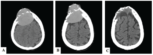
44-year-old female patient presented to our facility with 2 week history of headaches and massive mass located in the right forehead. She was diagnosed with multiple myeloma 5 years prior to presentation in outside hospital where she was initially treated with multiple lines including bortezomib and lenalidomide based multiple regimens in addition to 2 autologous stem cell transplants. Computed tomography (CT) brain scans (Figure 1) and Magnetic resonance imaging (MRI) scans (Figure 2) were representing extramedullary plasmacytoma. We treated patient with Dexamethasone and radiation therapy. Her mass had significant response to treatment, and later we referred patient to hospice secondary to deterioration in performance status. | ||||
Figures at a glance |
||||
|