Ahmet Okus1, Baris Sevinc2*, Omer Karahan3, Serdenay4 and Bekir Gurocak5
1General Surgery, Mevlana University Medical Faculty, Konya, Turkey
2General Surgery, Sarikaya Hospital, Yozgat, Turkey
3General Surgery, Meram Medical Faculty, Necmettin Erbakan University, Konya, Turkey
4General Surgery, Malazgirt State Hospital, Mus, Turkey
5General Surgery, Konya Training and Research Hospital, Konya, Turkey
- *Corresponding Author:
- Baris Sevinc
Sarikaya Hospital
General Surgery, Yozgat
Turkey
Tel: 00905054880511
E-mail: drbarissevinc@gmail.com
Received date: February 09, 2016; Accepted date: March 04, 2016; Published date: March 11, 2016
Keywords
Pilonidal disease; Flap; Primary closure; Oblique closure
Introduction
Pilonidal disease is a chronic inflammatory disease seen in the intergluteal region. The disease usually affects young adults and males. Hair is thought to be the main etiologic factor. Untreated disease affects life quality and causes morbidity [1-3].
There are many techniques for the surgical treatment of pilonidal disease. In surgical treatment after excision of the sinus tract totally, secondary healing, marsupialization, primary closure and flap reconstruction methods (Karydakis, Limberg flap, V-Y plasty etc.) can be chosen. However, there is no consensus on which is the best method.
The aim of this study is to compare the results of tension free primary closure (TPC), Limberg flap (LF) and oblique excision and primary closure (OC) in treatment of pilonidal sinus disease.
Material and Methods
Permission from Konya University Meram Medical Faculty Clinical Research Ethical Committee was taken. The groups were planned to be for 50 patients. An informed consent form was signed by all the patients. The patients were divided into 3 groups (tension free primary closure, Limberg flap, oblique excision and primary closure) by closed envelop method. The envelopes were opened at the operation room just before the surgery.
All the operations were performed under spinal anesthesia, in prone position. The sacral and gluteal regions were shaved at the operation day. Preoperative antibiotics and bowel preparation were not needed in any patients.
The patients, not suitable for OC (disseminated gluteal disease or perianal pilonidal sinus disease) were not included in the study. The remaining patients above 18 years of age were included into the study.
LF was performed in its classical form. The sinus tract was totally excised and a Limberg flap including skin and subcutaneous tissue was prepared from the right side. The subcutaneous tissue was closed by 2/0 polyglactine material as two folds separated sutures.
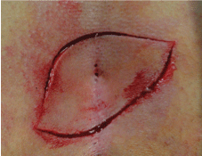
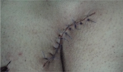
TPC; the sinus tract was totally excised with elliptical incision. Than subcutaneous tissue was released 2-3 cm in both sides. For OC the excision was performed with an oblique incision that includes the whole sinus tract (Figure 1). To prevent the tension at the wound, subcutaneous tissue was released as in TPC (Figure 2). In both procedures, subcutaneous tissue was sutured two folds by 2/0 polyglactine (Dogsan, Surgical Suture Materials, Turkey). Vacuumed drains were used for all the patients and the skin was closed with mattress sutures by 3/0 polypropylene (Dogsan, Surgical Suture Materials, Turkey). All the patients were discharged from the hospital at the first postoperative day. Drainage tubes were removed when the drainage was under 20 cc/24 hours. The patients were called to outpatient clinic and the drains were removed by the operating surgeon. Sutures were removed at the postoperative 14th day.

Figure 1: Ovarioles of the females emerged from control(a) and treated with different combinations of essential oils (b-h). Gr (Germarium),Vt (Vitellarium), VS (Vacant Space), LO (Lateral Oviduct).

Figure 2: The incision of oblique closure after the skin was sutured with 3/0 polypropylene.
The patients were called for follow up at the postoperative 14th day, 1st, 3rd and 6th months and yearly after.
The patients were examined and the findings were recorded at the outpatient clinic. At the follow up visits, drainage period, wound infection, wound dehiscence, seroma formation and painless sitting times were recorded.
For analysis of the data IBM SPSS statistics Version 20 was used. Results were presented as mean ± standard deviation. Students T test and Chi square tests were used where available and a p value lower than 0.05 was accepted as significant.
Results
At this stage there were 13 patients in LF group, 11 in OC group and 19 in TPC group. Due to the randomization method (closed envelope) the groups are not equal in number. The mean age of the patients was 28.5 ± 9.7. The demographic data were presented in Table 1.
| |
n |
Age (mean ± sd) |
Gender (M/F) |
| Limberg Flap (Group I) |
13 |
26.6 ± 8.3 |
09-Apr |
| Tension free primary closure (Group II) |
19 |
28.6 ± 8.9 |
18-Jan |
| Oblique closure (Group III) |
11 |
30.7 ± 12.9 |
06-May |
| Total |
43 |
28.5 ± 9.7 |
33/10 |
Table 1: Demographic data of the patients according to groups.
There was no significant difference in between the groups in terms of age, mean hospital stay, follow up time, body mass index and wound infection rate. The mean follow up time was 4.7 ± 1.3 months.
Wound infection was defined as; suppuration, heat increase and erythema around the wound. There was wound infection only in one patient in TPC group and it resolved by oral antibiotic therapy.
There was no seroma formation in LF and OC groups. However seroma formation was seen in 4 (21%) patients in TPC group. Seroma was drained once in 3 patients and twice in one patient. In none of those patients wound dehiscence was seen.
The mean operation time was significantly longer (55 minutes) in LF group. The mean follow up time, drainage period, painless sitting time, body mass index and operation time were presented in Table 2.
| |
Follow up time (months) |
Drainage period (days) |
Closet sitting time (days) |
Painless sitting time (days) |
Body Mass Index |
Operation time (minutes) |
| Limberg flap (Group I) |
5.1 ± 1.5 |
4.4 ± 1.2 |
10.9 ± 3.3 |
10.6 ± 3.3 |
24.1 ± 3.4 |
55 ± 11.5 |
| Tension free primary closure (Group II) |
4.1 ± 1.2 |
7.5 ± 2.3 |
7.5 ± 3.9 |
7.8 ± 3.2 |
25.3 ± 2.7 |
46.8 ± 6.9 |
| Oblique closure (Group III) |
5.3 ± 1 |
3.5 ± 0.9 |
11.3 ± 3.1 |
12.4 ± 2.7 |
24 ± 2.9 |
43.1 ± 9.8 |
| Total |
4.7 ± 1.3 |
5.6 ± 2.5 |
9.5 ± 3.9 |
9.9 ± 3.6 |
24.6 ± 3 |
48.3 ± 10.1 |
| p |
0.23 |
0.001 |
0.008 |
0.001 |
0.3 |
0.009 |
| The data was presented as mean ± standard deviation. |
Table 2: Follow up parameters of the groups.
Drainage period was 7.5 days in TPC group and it was significantly longer than other groups (p = 0.001). There was no difference in between OC and LF groups in terms of painless sitting time and back to work time. However, closet sitting time and painless sitting time was significantly shorter in TPC group (p = 0.008, p = 0.001, respectively).
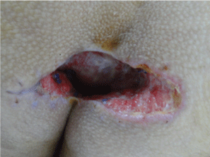
There was no wound dehiscence in LF and TPC groups. However, in OC group there was wound dehiscence in 10 of 11 (90.9%) patients; partial in 3 and complete dehiscence in 7 patients. The mean dehiscence time was 8.2 days. A patient with a non-healing wound after 2 months of follow up was re-operated (Limberg flap reconstruction) (Figure 3).

Figure 3: The wound that wasn’t closed after 3 months of follow up.
After this early evaluation, patient inclusion to the OC group was stopped. The study is still continuing with the other two groups.
Discussion
Pilonidal sinus disease is a chronic inflammatory disease, affecting young man. Recently, pilonidal sinus is accepted to be an acquired disease. The main etiologic factor is the hair and there are some other factors that ease the entrance of the hair [3]. There are so many studies evaluating the etiological factors of pilonidal sinus disease. In those studies, body mass index, gender, hair density, sitting time, shower frequency, local hygiene and family history are accepted as risk factors [4-8].
Karydakis reported his own method with no scar tissue at the midline and eliminating deep sulcus with a recurrence rate of lower than 1% [3]. Then, the main principle of the surgical treatment became elimination of the deep sulcus and leaving no scar tissue at the midline.
Therefore, many flap reconstruction methods are used in the surgical treatment of pilonidal disease like Limberg flap and V-Y flap [3,9,10]. It was reported that local hygiene and laser epilation of this region can reduce the recurrence rate [11,12].
Limberg flap reconstruction is a frequently used method with a recurrence rate lower than 5% [13-15]. However, recent studies have showed tension free primary closure and primary closure itself can be as successful as other methods [14,16,17]. Tension free primary closure eliminates the deep sulcus however, leaves scar tissue at the midline. It has no wound tension, can be performed easily and has good cosmetic results. It is suggested that the main factors to prevent the recurrences in tension free primary closure were local hygiene and tension free healing side [17].
In this study, the results of Limberg flap, tension free primary closure and oblique primary closure were compared.
One of the important aims of this study was to determine the importance of midline incision. In the literature there are limited studies abut oblique excision and primary closure. The largest study belongs to Mentes et al. [18]. They performed oblique excision and primary closure in 493 patients. They did not release the subcutaneous tissue and closed the wound with 0 polypropylene mattress sutures. They reported the recurrence rate as 5.6% at 18 months follow up. They reported the method to be useful as being easy, having shorter healing time and leaving no scar tissue at the midline. They reported 1.2% wound infection and 1% wound dehiscence rate. In the recent study there is 90.9% wound dehiscence rate in OC group. In our study OC was performed as tension free, however the difference with Mentes et al. is amazing. Moreover, in two studies by Akinci et al., limited asymmetrical elliptical incision has wound dehiscence rate of 1.8% in 112 patients and 4% in 24 patients [10,19]. Although, in those studies subcutaneous tissue was released to create a tension free healing side, the difference is still remarkable.
This study shows that, an oblique incision, different from Langer lines and body axis is a bad choice. There was wound dehiscence in almost all patients. According to this result OC group was excluded from the study.
There are many studies showing successful treatment of pilonidal disease with asymmetrical closure preventing midline scar tissue [10,19]. In all those studies wound side were released to prevent tension. The similar results of tension free primary closure and asymmetrical closure may be related to a tension free healing side. For this study, inclusion of another group as asymmetrical closure or Karydakis flap may make it more valuable. For prevention of postoperative pain and wound dehiscence tension free healing side should be the main principle [19]. As the study is still continuing, the recurrence rates were not evaluated.
As conclusion, the main goal of surgical treatment of pilonidal sinus disease should be local hygiene and tension free healing side. Oblique excision and primary closure is a bad choice in treatment of pilonidal disease.
9051
References
- McCallum I, King PM, Bruce J (2007) Healing by primary versus secondary intention after surgical treatment for pilonidal sinus. Cochrane Database Syst Rev 17: CD006213 PMID: 17943897.
- Alemderoglu K, Akçal T, Burga D (2003) Pilonidal disease. Diseases of the rectum and the anal region (1st edn.), Istanbul.
- Karydakis GE (1992) Easy and successful treatment of pilonidal sinus after explanation it’s causative process. Aust N Z J Surg 62: 385-389.
- Akinci OF, Bozer M, Uzunkoy A, Düzgun SA, Coskun K (1999) Incidence and etiological factors in pilonidal sinus among Turkish soldiers. Eur J Surg165: 339-342.
- Harlak A, Mentes O, Kilic S, Coskun K, Duman A, et al. (2010) Sacrococcygeal pilonidal disease: analysis o previously proposed risk factors. Clinics 65: 125-131.
- Conroy FJ, Kandamany N, Mahaffey PJ (2008) Laser depilation and hygiene: preventing recurrent pilonidal sinus disease. Journal of Plastic, Reconstructive Aesthetic Surgery61:1069-1072.
- Yilmaz Can MH, Joy MM, Yigit G Sharp O (2008) Sacrococcygeal pilonidal sinus Overweight young men, is associated with higher body mass index and skin color. Colon Rectum Haste Derg 18: 14-20.
- Sondenaa K, Andersen E, Nesvik I, Soreide JA (1995)Patient characteristics and symptoms in chronic pilonidal sinus disease. Int J Colorectal Dis 10: 39-42.
- Krand O, Yalt T, Berber I, Kara VM, Tellioglu G (2009) Management of pilonidal sinus disease with obliqueexcision and bilateral gluteus maximus fascia advancing flap: result of 278 patients. Dis Colon Rectum 52:1172-1177.
- Akinci OF, Coskun A, Uzunkoy A (2000) Simple and effective surgical treatment of pilonidal sinus: asymmetric excision and primary closure using suction drain and subcuticular skin closure. Dis Colon Rectum 43:701-706.
- Conroy FJ, Kandamany N, Mahaffey PJ (2008) Laser depilation and hygiene: preventing recurrent pilonidal sinus disease. Journal of plastic, Reconstructive Aesthetic Surgery 61: 1069-1072.
- Odili J, Gault D (2002) Laser depilation of the natal cleft-an aid to healing the pilonidal sinus. Ann R Coll Surg Engl84: 29-32.
- Akça T, Çolak T, Ustunsoy B, Kanik A, Aydin S (2005) Randomized clinical trial comparing primary closure with the Limberg flap in the treatment of the primary sacrococcygeal pilonidal disease. British J Surg 92: 1081-1084.
- Muzi MG, Milito G, Cadeddu F, Nigro C, Andreoli F,et al. (2010)Randomized comparison of Limberg flap versus modified primary closure for the treatment of pilonidal disase. The American Journal of Surgery 200: 9-14.
- Topgul K, Ozdemir E, Kiliç K, Okbayir H, Ferahkose Z (2003) Long-term result of Limberg flap procudere for treatment of pilonidal sinus: a report of 200 cases. Dis Colon Rectum 46: 1545-1548.
- Mahdy T (2008) Surgical treatment of the pilonidal disease: Primary closure or flap reconstruction after excision. Disease Colon Rectum 51: 1816-1822.
- Okus A, Sevinc B, Karahan O, Eryilmaz MA (2012)Comparison of Limberg Flap and Tension-Free Primary Closure During Pilonidal Sinus Surgery. World J Surg 36:431-435.
- Mentes O, Bagci M, Bilgin T, Coskun I, Ozgul O,et al. (2006)Management of pilonidal sinus disease with obliqueexcision and primary closure: results of 493 patients.Dis Colon Rectum 49:104-108.
- Akinci OF, Coskun A, Ozgonul A, Terzi (2006) A Surgical treatment of complicated pilonidal disease: limited separate elliptical excision with primary closure.Colorectal Dis.8:704-709.


