Labuda Craig S1 Luu Sylvester2 Thompson Kaylyn3* and Berry-Cabán Cristóbal3
1Musculoskeletal Radiology, Department of Radiology, Womack Army Medical Center, Fort Bragg, NC, USA
2Western University of Health Sciences, College of Osteopathic Medicine, Pomona, CA, USA
3Department of Clinical Investigation, Womack Army Medical Center, Fort Bragg, NC, USA
- *Corresponding Author:
- Thompson Kaylyn
Department of Clinical Investigation
Womack Army Medical Center
Fort Bragg, NC, USA
E-mail: krthompson0623@email.campbell.edu
Pectoralis major muscle injuries occur most commonly among men between the ages of 20 and 39 and often take place during weightlifting activities while the arm is contracted during external rotation and/or extension [1,2]. Pectoralis major injuries may present as an audible pop, tearing sensation, instantaneous pain and weakness with swelling and ecchymosis in the anterior axillary fold and lateral chest, and upper arm weakness with adduction [1,3-9]. This type of injury is relatively uncommon and accurate diagnosis may be overlooked if not correctly assessed. Diagnostic imaging confirmation is often obtained with magnetic resonance imaging (MRI). Most pectoralis major injuries in the active-duty military population occur during bench-press exercises, though a subset of injuries occur during airborne operations.
Keywords
Pectoralis major tendon rupture, Soldiers, Paratrooper
Introduction
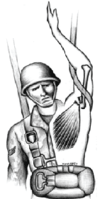
Pectoralis major muscle injuries occur most commonly among men between the ages of 20 and 39 and often take place during weightlifting activities while the arm is contracted during external rotation and/or extension [1,2]. Pectoralis major injuries may present as an audible pop, tearing sensation, instantaneous pain and weakness with swelling and ecchymosis in the anterior axillary fold and lateral chest, and upper arm weakness with adduction [1,3-9]. This type of injury is relatively uncommon and accurate diagnosis may be overlooked if not correctly assessed. Diagnostic imaging confirmation is often obtained with magnetic resonance imaging (MRI). Most pectoralis major injuries in the active-duty9 military population occur during bench-press exercises, though a subset of injuries occur during airborne operations. Pectoralis tendon injuries in paratroopers typically occur during the opening jolt of the parachute or when the arm becomes entangled in the static line of the parachute upon exit causing excessive pull, hyperabduction, and external rotation of the limb (Figure 1) [10].10 Static line injuries account for the second largest number of injuries among the airborne soldiers [11-13].

Figure 1: Illustration of the opening shock injury of the paratrooper. Excessive traction. Hyperabduction, and external rotation of the arm caused muscle rupture.
We report a case of an active duty male soldier with a ruptured pectoralis major tendon from a static line entanglement during an airborne training exercise.
Case Description
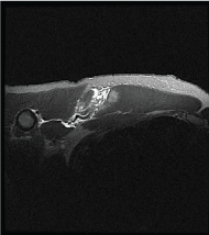
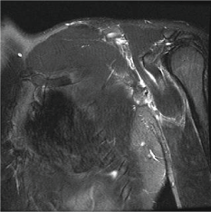
During a routine airborne training exercise, a 20-year-old active duty male soldier serving in a military airborne operations unit entangled his left wrist and forearm in the static line upon exiting the aircraft. The restrained left arm underwent hyperflexion and external rotation. After a period of entanglement, the left upper extremity was ultimately released and the remaining airborne operation was uneventful. Once on the Drop Zone the patient reported severe left upper arm, axilla, and lateral chest wall pain. The patient presented to the medical clinic for evaluation with 8 out of 10 pain and inability to perform daily strenuous activities with his left arm. Physical examination findings revealed left anterior upper outer chest wall edema and ecchymosis and visual defect at the left axillary fold. An acute pectoralis major tear was suspected. The left shoulder radiograph was normal, though subsequent MRI examination revealed complete a sternocostal head pectoralis major tendon tear with retraction and partial thickness muscle tear (Figures 2 and 3).

Figure 2: Full width sternocostal head pectoralis major tendon tear with tendon retraction and partial thickness muscle laceration.

Figure 3: Retracted pectoralis major tendon and surrounding edema.
Surgical repair was performed approximately one month after the traumatic incident using a deltopectoral surgery. Blunt dissection exposed the bicipital groove and the retracted tendon was identified. Three whipstitch sutures secured the tendon that was subsequently secured to three pec buttons. These buttons were then anchored to the lateral wall of the bicipital groove through three pilot holes.
Simple sling was applied immediately post-operative. Rehabilitation at weeks 1 and 2 involved scapular stabilizer muscle sets, passive range of motion (ROM) with forward flexion to 90 degrees and passive abduction ROM to 60 degrees, and pendulum exercises. Physical therapy sessions progressed to active ROM to point of first resistance, gentle towels stretch, upper body ergometer, and internal and external strengthening with light theraband. A temporary profile was adjusted during the rehabilitation process to protect from re-injury. Unrestricted airborne, physical fitness testing, heavy bench press exercises, other demanding occupational activities were reinstated at approximately 12 months postoperative.
Discussion
Pectoralis major injuries are not uncommon in the active duty population.2 There were 43 surgically repaired pectoralis major tears at Womack Army Medical Center between 2012 to 2014 with 55 percent of these injuries occurring during weightlifting exercises and 23 percent (second most common) occurred from static line injuries during airborne operation maneuvers.
Radiographs are routinely obtained to exclude acute bone injury [14]. The most common type of diagnostic cross sectional imaging performed for this type of injury is magnetic resonance imaging. MRI can accurately identify location and extent of injury, and assist surgical planning for appropriate candidates. Classification of pectoralis major tears was recently described by Chiavaras, et al. [15] When reviewing an MRI examination, the axial T2 fat suppression and axial T1 sequences offer exquisite anatomic detail of both sternal and clavicular head, musculotendinous junction, tendon and distal tendon attachment. Pectoralis tendon injuries most commonly occur at the myotendinous junction or tendon, with proximal muscle tears being less common. MRI findings of soft tissue edema adjacent to the humerus or periosteal edema are often associated with a distal tendon tear or avulsion.
Successful surgical repair and full return to active duty activities in the soldier was contributed by accurate clinical diagnostic assessment, descriptive MRI interpretation of location and extent of injury for proper surgical planning, and timely surgical intervention with aggressive rehabilitation.
Timely diagnostic imaging and accurate interpretation in conjunction with rapid surgical evaluation and repair allows for optimal successful patient outcome [2,16].
Acknowledgements
We would like to acknowledge Dr. Mahmut Komurcu (Author) and Mr. Tuna Ferit (Illustrator) for the use of the illustration in Figure 1.
Disclaimer
The views expressed herein are those of the author(s), and do not reflect the official policy of the Department of the Army, Department of Defense, or the United States government. This study used no outside funding of any kind.
7076
References
- Provencher MT, Handfield K, Boniquit NT, Reiff SN, Sekiya JK, et al. (2010) Injuries to the pectoralis major muscle: diagnosis and management.Am J Sports Med 38: 1693-1705.
- Aärimaa V, Rantanen J, Heikkilä J, Helttula I, Orava S (2004) Rupture of the pectoralis major muscle.Am J Sports Med 32: 1256-1262.
- Alho A (1994) Ruptured pectoralis major tendon. A case report on delayed repair with muscle advancement.ActaOrthopScand 65: 652-653.
- Bakalim G (1965) Rupture of the pectoralis major muscle: a case report. ActaOrthopaedica36:274-279.
- Petilon J, Carr DR, Sekiya JK, Unger DV (2005) Pectoralis major muscle injuries: evaluation and management.J Am AcadOrthopSurg 13: 59-68.
- Potter BK, Lehman RA Jr, Doukas WC (2004) Simultaneous bilateral rupture of the pectoralis major tendon. A case report.J Bone Joint Surg Am 86-86A: 1519-21.
- Schepsis AA, Grafe MW, Jones HP, Lemos MJ (2000) Rupture of the pectoralis major muscle. Outcome after repair of acute and chronic injuries.Am J Sports Med 28: 9-15.
- Arciero RA, Cruser DL (1997) Pectoralis major rupture with simultaneous anterior dislocation of the shoulder.J Shoulder Elbow Surg 6: 318-320.
- Mackenzie DB (1981) Avulsion of the insertion of the pectoralis major muscle. A case report.S Afr Med J 60: 147-148.
- Komurcu M, Yildiz Y, Ozdemir MT, Erler K (2004) Rupture of the pectoralis major muscle in a paratrooper.Aviat Space Environ Med 75: 81-84.
- Bricknell MC, Craig SC (1999) Military parachuting injuries: a literature review.Occup Med (Lond) 49: 17-26.
- Knapik JJ, Steelman R, Grier T, Graham B, Hoedebecke K, et al. (2011) Military parachuting injuries, associated events, and injury risk factors.Aviat Space Environ Med 82: 797-804.
- Potter RN, Gardner JW, Deuster PA, Jenkins P, McKee K Jr, et al. (2002) Musculoskeletal injuries in an Army airborne population.Mil Med 167: 1033-1040.
- Simonian PT, Morris ME (1996) Pectoralis tendon avulsion in the skeletally immature.Am J Orthop (Belle Mead NJ) 25: 563-564.
- Chiavaras MM, Jacobson JA, Smith J, Dahm DL (2015) Pectoralis major tears: anatomy, classification, and diagnosis with ultrasound and MR imaging.Skeletal Radiol 44: 157-164.
- Bak K, Cameron EA, Henderson IJ (2000) Rupture of the pectoralis major: a meta-analysis of 112 cases.Knee Surg Sports TraumatolArthrosc 8: 113-119.


