Deborah Repullo Jennen*, Eric Lebrun, Jean Lemaitre, Didier Hossey and Alexis Therasse
Department of Visceral Surgery; CHU Ambroise Paré Mons, Belgium
*Corresponding Author:
Deborah Repullo Jennen
Chirurgie des tissus mous
CHU Ambroise Paré
Boulevard Kennedy 2
7000 Mons, Belgium
E-mail: Deborah.Repullojennen@hap.be
A 54 year-old woman with a history of giant type I hiatal hernia, severe asthma, and obstructive sleep apnea presented at the emergency room with epigastric pain. Abdominal CT scan showed a giant intrathoracic hiatal hernia and a massive pneumopericardium together with an image compatible with peptic ulcer perforation. Within 2 hours a surgical peptic ulcer repair was performed by thoracotomy in the 8th rib space and covered by a pericardial fat patch.
Case Report
The patient was not under proton pump inhibitors and was known to follow a chronic methylprednisolone treatment for her asthma. A Nissen procedure was realized 4 years ago in another institution with intrathoracic migration afterwards due to short oesophagus. Upon examination the patient presented with hypotension (systolic pressure < 80 mmHg), epigastric pain and decreased heart sounds. No defense, no fever and no abnormal pulmonary examination were evidenced at the emergency department.
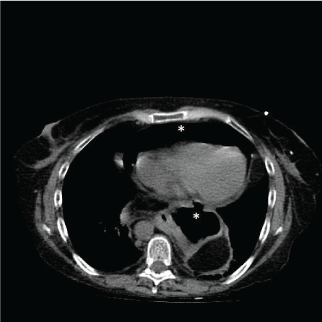
Electrocardiogram and cardiac ultrasonography were noncontributive. Abdominal CT scan showed a giant intrathoracic hiatal hernia and a massive pneumopericardium together with an image compatible with peptic ulcer perforation. The probable diagnosis of a peptic ulcer perforated into the pericardium was then established. Within 2 hours a surgical peptic ulcer repair was performed by thoracotomy in the 8th rib space closing the perforated ulcer in two layers and covering it by a pericardial fat patch. Two right thoracic drains were left in place and the stomach decompressed by a naso-gastric tube.
The patient was covered by proton pump inhibitors at high dose. During early postoperative care the patient developed bilateral inferior lobar atelectasis and a right pleural effusion which resolved with thoracic drainage and intensive respiratory physiotherapy. Drains were removed at day 11 and 12 A contrast oesogastroduodenography was performed before oral feeding. The patient was discharged from hospital 3 weeks after the operation.
Discussion
This case presents 3 concomitant rare clinical situations: 1) peptic ulcer perforation of hiatal hernia, described as iatrogenic or associated to paraoesophageal hernias [1-4] or during postpartum [5]; 2) fistulisation of a perforated ulcer into the pericardium which has been described among surgical complications after Nissen fundoplication [6] and only twice over the latest 15 years with a spontaneous perforation [7,8]; and 3) pneumopericardium, described as a result of blunt thoracic injuries [9] and among complications of gastric bypass surgery [10,11] Outcomes of these complications are poor and there are currently no guidelines for their management (Figure 1).

Figure 1: Image of peptic ulcer perforation and pneumopericardium.
The use of noninvasive complementary examinations and an early surgical treatment probably contributed to the positive outcome of our patient. Therefore it seems necessary to discuss the importance of an early multidisciplinary approach and whether proton pump inhibitors should be systematically prescribed to patients presenting a hiatal hernia. This seems particularly beneficial in some particular cases such as patients with a history of surgical procedures like Nissen fundoplication or chronic corticosteroids intake which could mitigate the clinical symptoms of a peptic ulcer leading to a delay in the diagnostic of certain complications.
Acknowledgements
We would like to thank the rest of the surgical team who actively contributed to the positive outcome of this patient, as well as the non-surgical teams: both the radiology and pneumology departments of CHU Ambroise Paré.
7115
References
- Laski D, Lukianski M, Dubowik M, Pawlaczyk R, Zurek W, et al. (2014) Perforation of benign peptic ulcer in hiatal hernia into the pericardium, resulting in pneumopericardium.Endoscopy 46 Suppl 1 UCTN: E423.
- Shiwani MH, Thornton MP (2008) Supradiaphragmatic perforated duodenal ulcer in a giant hiatus hernia.Can J Surg 51: E89-90.
- Ekelund M, Ribbe E, Willner J, Zilling T (2006) Perforated peptic duodenal ulcer in a paraesophageal hernia--a case report of a rare surgical emergency.BMC Surg 6: 1.
- Pol RA, Wiersma HW, Zonneveld BJ, Schattenkerk ME (2008) Intrathoracic drainage of a perforated prepyloric gastric ulcer with a type II paraoesophageal hernia.World J EmergSurg 3: 34.
- FilippoLococo, Alfredo Cesario, Elisa MeacciIntrathoracic. Gastric perforation: a late complication of an unknown postpartum recurrent hiatal hernia. Interactive CardioVascular and Thoracic Surgery 15: 317-319.
- Murthy S, Looney J, Jaklitsch MT (2002) Gastropericardial fistula after laparoscopic surgery for reflux disease.N Engl J Med 346: 328-332.
- Bruhl SR, Lanka K, Colyer WR Jr (2009) Pneumopericardialtamponade resulting from a spontaneous gastropericardial fistula.Catheter CardiovascInterv 73: 666-668.
- Riepe G, Braun S, Swoboda L (1998) [Perforation into the heart--a rare complication of stomach ulcer in hiatal hernia].Chirurg 69: 475-476.
- Haan JM, Scalea TM (2006) Tension pneumopericardium: a case report and a review of the literature.Am Surg 72: 330-331.
- Dhillon A, Eltweri AM, Shah V, Bowrey DJ (2015) Gastropericardial fistula after Roux-en-Y bypass for reflux disease.BMJ Case Rep 2015.
- Huyskens J, Macken E, Schurmans J, Parizel PP, Salgado R (2014) A case of pneumopericardium as a late complication of gastric bypass surgery.Circulation 130: 1633-1635.


