Keywords
|
| Prostate cancer, Radiotherapy, Postradiation sarcoma |
Introduction
|
| Radiotherapy for prostate cancer can be associated with the development of second cancer. The majority are carcinomas while post-irradiation sarcomas are exceptional. We report the case of a 75 year-old Caucasian male who was diagnosed with a pelvic sarcoma eight years after external beam radiotherapy. |
Case Presentation
|
| A seventy-five year-old Caucasian male presented with rapidly progressive incontinence and obstipation eight years after high dose radiotherapy for prostate adenocarcinoma. |
| At the age of 67, the patient had been diagnosed with moderately differentiated localized T1c adenocarcinoma with a Gleason score of 6(3+3) using needle biopsy of the prostate. Pre-treatment Prostate-Specific-Antigen (PSA) had been 5.63 ng/ml (normal range < 4 ng/ml). |
| First, he underwent radiation therapy at 80 Gy on the and 54 Gy on the seminal glands. PSA nadir of 0.98 was achieved in four years after treatment, and no urinary or digestive toxicity were described. |
| Five years after irradiation, PSA had risen slightly and the patient began Leuproreline three years later, after the Positron Emission Tomography (PET) imaging had showed a localized prostate hypermetabolism and that PSA had reached 4.45 ng/ml. |
| New needle biopsy of the prostate revealed a Gleason score of 8(4+4) adenocarcinoma. |
| After one year of hormonal ablation therapy the patient showed obstructive symptomatology and septicemia. |
| On physical examination, he had pelvic and lumbar pain, constipation, substancial increase in nocturia and dysuria. |
| The patient received large-spectrum antibiotics and underwent derivation surgery with a colostomy. Transanal-guided biopsies were performed. PSA levels continued to be low at 1.79 ng/ml. |
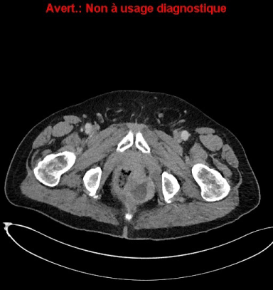
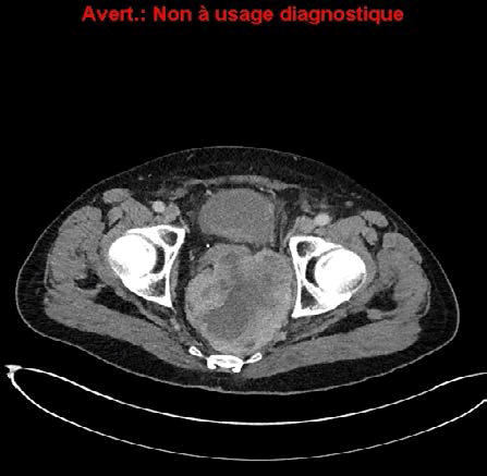
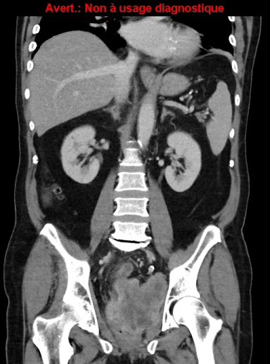
| Abdominal Computed Tomography (CT) scan revealed a huge heterogeneous mass measuring 10.5 × 10.7 cm. Thorax CT scan did not find lung dissemination (Figure 1). |
| Prostate high-grade sarcoma with rectum extension was suspected (Figures 2 and 3). |
| On histological examination, mitoses were easily found: 3/10 fields and there were multiple foci of necrosis. On immunohistochemistry, the tumor cells were positive for CD31 and negative for: cytokeratine AE1-AE3, CK7,CK5-6,CK20,EMA,P63,PSA,CDX2,PSO4S, chromogranine A,PS100,CD34,Actine, Desmine, Caldesmone, CD117,DOG1. |
| On the basis of these results, the tumor was classified as a highgrade (III) pleomorphic undifferentiated cell sarcoma, linked to radiotherapy. |
| Sarcomatoid sarcoma was excluded because of the absence of epithelial markers. The patient, due to the invasion of the rectum, was not eligible for surgery and received neoadjuvant based-Anthracycline chemotherapy. After three cycles of chemotherapy with DOXORUBICIN 50 mg/m2 every three weeks, central necrosis of the tumor was observed, but a metastasis in the liver appeared. |
| Chemotherapy was followed. After 6 cycles of chemotherapy, partial response was obtained. Tumor surgery and metastasectomy were planned. |
Discussion
|
| The first clinical association between exposure to radiotherapy and sarcoma was recognized in 1992 by Beck, in a description of bone sarcomas in patients who had received radiotherapy for tuberculous arthritis [1]. These tumors usually occur many years after exposure to radiation and are associated with a poor prognosis [2]. Post-irradiation bone sarcomas are more common than soft tissue sarcomas. Radiation-induced sarcomas are rare tumors, more malignant that prostatic adenocarcinoma, representing less than 0.2% of all irradiated patients [3]. Local symptoms are the most frequent presentation. In our case the patient presented subocclusion symptoms; that lead to diagnosis [4]. |
| CT scan is usefull for the diagnosis, because it will help to study locoregional and distant spread of the disease. Nevertheless the definitive diagnosis is established through biopsy. Several publications suggest that there is an increased risk of second cancers after radiotherapy or brachytherapy [5]. |
| Cahan et al. established in 1948 criteria to diagnose a postirradiation neoplasm: localization of the tumor, latent period over than four years, completely different histological between the two lesions, any genetic predisposition for tumor [6]. Our patient had no familial history that could suggest a hereditary genetic disorder. Brenner et al. found that 1 of 250 irradiated patients for prostate cancer would develop secondary cancer after 5 years, while after 10 years 1 of 70 patients would [7]. The second cancers appear in the radiation field, adjacent to it or in distant sites. These authors reported the high risk for developing pelvic sarcoma after radiotherapy that was 85% (IC95% [15-201%]). |
| For patients with bulky disease who may be at risk for positive surgical margins, small series have demonstrated that neoadjuvant doxorubicin and cisplatin can produce significant tumor necrosis [8]. Experimental studies suggest that intensity modulated radiation therapy may be associated with up to three times higher risk of secondary malignancy [9]. |
| There are few cases of radiation-induced prostatic sarcoma reported in the medical literature. Due to the increased-life expectancy with improved survival in cancer patients, the incidence is expected to be higher. |
Conclusion
|
| Sarcoma of the prostate is a rare and aggressive malignancy that can develop many years after treatment for prostate adenocarcinoma. |
| In addition to long-term surveillance for disease recurrence, there should be suspicion for development of second cancer when obstructive symptoms occur without biochemical relapse, particularly when the patient has a history of pelvic irradiation. |
Figures at a glance
|
 |
 |
 |
| Figure 1 |
Figure 2 |
Figure 3 |
|
| |
| |


