Key Words
QUS, Stiffness index, Osteopenia, Osteoporosis, T-score, Young Saudi Females.
Introduction
The bone is characterized by its rigidity and hardness to provide structural support for the body protection of vital organs, an environment for marrow (both blood forming and fat storage) and to act as a mineral reservoir for calcium homeostasis in the body [1].
Although dual-energy x-ray absorptiometry (DXA) and quantitative computed tomography (OCT) are two non-invasive procedures being reliable and accepted tests for osteoporosis in clinical practice [2] limited availability and the ionization nature of both tools have stimulated interest in safer, portable, and less expensive techniques to identify those at risk of osteoporotic fracture.
Quantitative ultrasound (QUS) has been introduced as an indirect assessment of bone quality [3-4] with the technique being attractive due to the non-ionizing nature of the procedure, lower cost and portability of the device [5]. The theory of QUS is based on the principle that bone as a porous material will absorb, scatter and transmit sound wave dependent on stiffness, density and volume of the material [6-7]. Both sound attenuation and the sound velocity are combined to form the stiffness index used in commercial units. Parameters measured by QUS were found to correlate with trabecular volume and thickness which manifest strength and load-bearing capacity [8].
Both in-vitro and in-vivo studies showed that QUS and bone densitometry results are highly correlated as long as they are measured along a single direction and at the same location [9-10].
Potential clinical use of QUS devices includes diagnosis of osteoporosis, fracture risk assessment and monitoring of skeletal changes associated with disease prognosis and treatment. Osteoporosis is a systemic skeletal disorder, characterized by low bone mass, microarchitectual deterioration of bone tissue and an increase in bone fragility and susceptibility to fracture [1,11].
The aim of the study is to evaluate and measure stiffness index in young university aged Saudi females.
Materials and methods
Study Design
101 young female adults were enrolled in the study (age range 20–24.9 years), following the distribution of the announcements of the study among young females. Informed consent was obtained from all subjects, and the study was approved by local research ethics committee.
Each subject was interviewed using a standardized questionnaire to collect information on life style, smoking habits, and level of physical activity in leisure time; coffee and tea consumption, illness and the use of medications. Pregnant females, females stating that they have any disease or taking any medication affecting their bone or suffering from any terminal illness were excluded from the study.
Age, body weight, height and body mass index (BMI) were recorded and QUS was applied to all subjects enrolled.
QUS measurements
Measurements were made using Lunar Achilles Insight TM - GE Healthcare which is a heel water-bath ultrasound system. It generates a band of frequencies from 200 to 600 kHz. Stiffness index (automatically calculated from broadband ultrasound attenuation and the speed of sound), T-score and Z-score were recorded using a standard protocol supplied by the manufacturer. Daily quality control was carried out using the quality phantom. At the time of the study, data collection could not be started unless quality control test is passed and measurements were conducted on the independent foot.
Statistical Analysis
Using SPSS software, One-way Anova was used to examine differences in Stiffness index between subjects with and without osteoporosis and osteopenia. Pearson Correlation Coefficient was used to examine the presence of an association between age, weight, height, BMI index and Stiffness index, T-score and Z-score values in the calcaneus.
Results
Age and anthropometric data of the subjects studied are presented in Table 1.
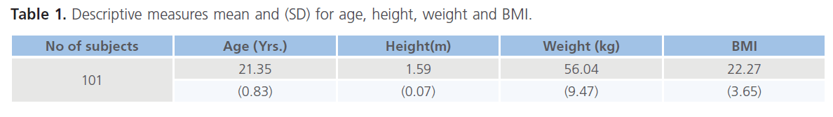
Table 1. Descriptive measures mean and (SD) for age, height, weight and BMI.
Of the 101 female subjects enrolled in the study, 3 subjects had T-score <-2.5 equivalent to the presence of osteoporosis in the calcaneus whereas 33 females had T-score (-1 to -2.5) suggestive of the presence of osteopenia within the calcaneus. 65 subjects had T-score > -1 equivalent to the absence of both osteopenia and osteoporosis Table 2.
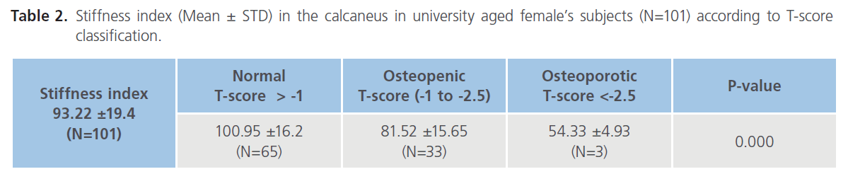
Table 2. Stiffness index (Mean ± STD) in the calcaneus in university aged female’s subjects (N=101) according to T-score classification.
Stiffness index (Mean ± STD) in the calcaneus according to T-score classification is tabulated in Table 2. Mean stiffness index in 3 subjects with a T-score <-2.5 (classified as osteoporotic) was equal 54.33 compared to 81.52 in 33 subjects with a T-score between -1 to -2.5 (classified as osteopenic) and a mean stiffness index value of 100.95 in 65 subjects with a T-score above -1 (considered as non-osteopenic nor osteoporotic). Results demonstrated a significant statistical reduction in stiffness index associated with a T-score equal to -1 and below in the calcaneus (P= 0.000).
The association between QUS parameters (stiffness index, T-score and Z-score) measured and age, weight, height, BMI index was investigated and tabulated in Table 3. A positive correlation were found between weight and the three QUS parameters (P < 0.01 for stiffness index) and (P < 0.05 for both T-score and Z-score) with no significant correlation between height and QUS parameters. The presence of correlation in weight and its absence in height with QUS parameters resulted in the presence of significant correlation between BMI index and stiffness index only (P < 0.05) as BMI index is a result of dividing weight (Kg) by height squared (m2).
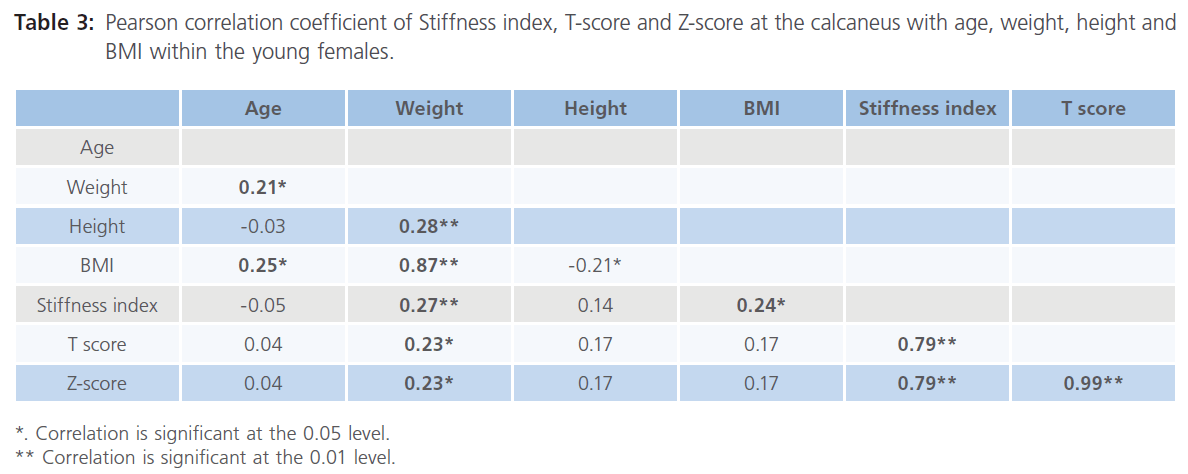
Table 3: Pearson correlation coefficient of Stiffness index, T-score and Z-score at the calcaneus with age, weight, height and BMI within the young females.
Discussion
Bone is a highly specialized support tissue which is characterized by its rigidity and hardness. In females and during adolescence up to 90% of peak bone mass is acquired by early twenties [12] so the female passes to adulthood with the skeleton growing both in size and density with more bone deposited than withdrawn [13]. When this balance tips toward excessive resorption, bones weaken (osteopenia) and over time can become brittle and prone to fracture (osteoporosis).
Peak bone mass which occur during puberty which play an important role in the determination of osteoporotic fracture risk can be measured by specific tools. World Health Organization (WHO) defines bone mineral density as a tool in the assessment of bone strength in clinical practice [14]. Limited availability and the ionization nature of DXA and CT have stimulated interest in safer, portable, and less expensive techniques to identify those at risk of osteoporotic fracture.
QUS in bone has been proposed to provide information related to bone structure [6]. It is a simple inexpensive and non-invasive technique which does not contain any radiation. QUS was suggested as a tool to predict osteoporosis [3], a disorder characterized by low bone mass and an increase in bone fragility and susceptibility to fracture [1,11].
The aim of the study was to provide information about stiffness index measured by QUS in young Saudi females as a parameter related to bone strength and to establish a baseline measurement that would help later to identify changes in bones related to osteopenia and osteoporosis in Saudi Arabia. Our result showed a mean stiffness index equal to 93.22±19.4 in the whole sample, comparison with previous published data with similar age where not possible, a comparison with other age groups post-pubescent girls, mean age 14.9 ±0.6 years) and premenopausal Japanese population (40-49years) found similarities despite the age differences between groups [15-16]. Such similarity in stiffness indices in the calcaneus may arise from either ethnic difference in bone density as it is known that the Asian have smaller bone frame than Caucasians. The similarity in stiffness index between groups does not provide an explanation to the increase in bone density which occur at early twenties as peak bone is reported to occur at twenty years and continue to a premenopausal age [12].
When the Stiffness index data were stratified according to T-score values assigned for osteopenia (-1 to -2.5) and osteoporosis (≤-2.5), a significant reduction in stiffness index was found in 36 subjects out of 101 females (P=0000). Although, the sample included in the study stated that they did not suffer from any disease nor taking any medication affecting their bone, a reduction in stiffness index in more than third of the sample studied represent an emerging problem in Saudi Arabia and would require further investigation.
Mean Stiffness index in the neither non osteopenic nor osteoporotic group was found to be 100.95 ±16.2 which is higher than both previous studies in the post-pubertal girls and premenopausal females [15-16], which may reflect an improvement in bone strength at the beginning of the third decade of life. Further studies on wider age range are required to examine such suggestion.
The presence of T-score below -1 in more than a third (≈33% with T-score -1 to -2.5 and the rest 3% with a T-score ≤-2.5) of the sample studied suggest that the presence of osteopenia and osteoporosis is not confined to the postmenopausal community in Saudi Arabia [17] but it have earlier origins. The study demonstrated the need to examine both genetic and environmental contribution to bone mass, including the effect of diet and the absence of a program for physical activities for the Saudi females which resulted in the presence of low levels of mechanical loading affecting growing bone. The ability to use QUS to predict fracture and to determine who might be most beneficial from preventive measures will be useful [16], due to the increase incidence of osteoporosis and osteopenia within the region [17]. Studies both in-vitro and in-vivo demonstrated that QUS and bone densitometry results are highly correlated provided that they are measured along a single direction and at the same location [9-18], although further studies are required to correlate QUS in the calcaneus and BMD measurements in the spine and femur areas by DXA.
The positive correlation found between BMI and Stiffness index is contributed to the effect of weight and not height; results from this study are consistent with previous reports, where increasing weight is known to be associated with higher bone density [2,19]. Such findings suggest the importance of considering body weight in the evaluation of patients in relation to the diagnosis of osteoporosis [20].
The study demonstrates the ability to use QUS to examine bone quality in the calcaneus of the young adult females, with a reduction in stiffness index – corresponding to osteopenia and osteoporosis- in a third of the sample studied. Whether the reduction in the calcaneus area is equivalent to a reduction in the spine and femur require further investigation to determine whether the etiology of low QUS values is different from that of low bone mass and will open the possibility for QUS to be used as a screening tool in osteoporosis.
In conclusion, the present study shows that stiffness index values for the young Saudi adult females not suffering from osteoprosis or osteopenia are higher than values obtained from the premenstrual Caucasians and premenopausal Japanese females. A reduction in the stiffness index in the third of the sample was found corresponding to the presence of osteopenia. Weight has a positive relationship with bone mineral density.
Decleration of intrest
The Author reports no conflicts of interest and is responsible for the content and writing of the paper.
333
References
- Frost, HM. The pathomechanics of osteoporosis. Clin Orthop Relat Res. 1985; 200: 198-225.
- Mazess, RB., Barden, HS. Measurement of bone by dual-photon absorptiometry (DPA) and dual-energy X-ray absorptiometry (DEXA). Ann Chir Gynaecol. 1988; 77 (5-6): 197-203.
- Gregg, EW., Kriska, AM., Salamone, LM., Roberts, MM., Anderson, SJ., Ferrell, RE., Kuller, LH., Cauley, JA. The epidemiology of quantitative ultrasound: A review of the relationships with bone mass, osteoporosis and fracture risk. Osteoporos Int. 1997; 7 (2): 89-99.
- Njeh, CF., Boivin, CM., Langton, CM. The role of ultrasound in the assessment of osteoporosis: A review. Osteoporos Int. 1997; 7: 7-22.
- Gluer, CC. Quantitative ultrasound techniques for the assessment of osteoporosis: Expert agreement on current status. The International Quantitative Ultrasound Consensus Group. J Bone Miner Res. 1997; 12: 1280-1288.
- Langton, CM., Palmer, SB., Porter, RW. The measurement of broadband ultrasonic attenuation in cancellous bone. Eng Med. 1984; 13: 89-91.
- McKelvie, ML., Palmer, SB. The interaction of ultrasound with cancellous bone. Phys Med Biol. 1991; 36: 1331-40.
- Gluer, CC., Wu, CY., Jergas, M., Goldstein, SA., Genant, HK. Three quantitative ultrasound parameters reflect bone structure. Calcif Tissue Int. 1994; 55: 46-52.
- McKelvie, ML., Fordham, J., Clifford, C., Palmer, SB. In vitro comparison of quantitative computed tomography and broadband ultrasonic attenuation of trabecular bone. Bone 1989; 10: 101-104.
- Glüer, CC., Vahlensieck, M., Faulkner, KG., Engelke, K., Black, D., Genant, HK. Site-matched calcaneal measurements of broad-band ultrasound attenuation and single X-ray absorptiometry: do they measure different skeletal properties? J Bone Miner Res. 1992; 7 (9): 1071-9.
- Melton, LJ.3rd, Wahner, HW. Defining osteoporosis. Calcif Tissue Int. 1989; 45 (5): 263-4.
- Matkovic, V., Jelic, T., Wardlaw, GM., Ilich, JZ., Goel, PK., Wright, JK., Andon, MB., Smith, KT., Heaney, RP. Timing of peak bone mass in Caucasian females and its implication for the prevention of osteoporosis. Inferencefrom a cross-sectional model. J Clin Invest. 1994; 93 (2): 799-808.
- Bonjour, JP., Theintz, G., Buchs, B., Slosman, D., Rizzoli, R. Critical years and stages of puberty for spinal and femoral bone mass accumulation during adolescence. J Clin Endocrinol Metab. 1991; 73 (3): 555-63.
- World Health Organization (WHO). Assessment of fracture risk and its application to screening for postmenopausal osteoporosis: Report of a WHO Study Group, World Health Organ Tech Rep Ser 843. 1994. pp. 1-129.
- Arnett, MG., Lutz, B. Effects of rope-jump training on the oscalcis stiffness index of postpubescent girls. Med Sci Sports Exerc. 2002; 34 (12): 1913-9.
- Yoshimi, I., Aoyagi, K., Okano, K., Yahata, Y., Kusano, Y., Moji, K., Tahara, Y., Takemoto, T. Stiffness index of the calcaneus measured by quantitative ultrasound and menopause among Japanese women: The Hizen-Oshima Study. Tohoku J Exp Med. 2001; 195 (2): 93-9.
- Rouzi, AA., Al-Sibiani, SA., Al-Senani, NS., Radaddi, RM., Ardawi, MS. Independent predictors of all osteoporosisrelated fractures among heatlhy Saudi postmenopausal women: the CEOR Study. Bone 2012; 50 (3): 713-22.
- Waud, C., Lew, R., Baran, DT. The relationship between ultrasound and densitometric measurements of bone mass at the calcaneus in women. Calcif Tissue Int. 1992; 51: 415-418.
- Maalouf, G., Salem, S., Sandid, M., Attallah, P., Eid, J., Saliba, N., Nehme, I., Johnell, O. Bone mineral density of the Lebanese reference population. Osteoporosis Int. 2000; 11: 765-769.
- Kudlacek, S., Schneider, B., Peterlik, M., Leb, G., Klaushofer, K., Weber, K., Woloszczuk, W., Willvonseder, R. Normative data of bone mineral density in an unselected adult Austrian population. Eur J Clin Invest. 2003; 33: 332-339.


