Flyer

Health Science Journal

  • ISSN: 1108-7366
  • Journal h-index: 61
  • Journal CiteScore: 17.30
  • Journal Impact Factor: 18.23
  • Average acceptance to publication time (5-7 days)
  • Average article processing time (30-45 days) Less than 5 volumes 30 days
    8 - 9 volumes 40 days
    10 and more volumes 45 days
Awards Nomination 20+ Million Readerbase
Indexed In
  • Genamics JournalSeek
  • China National Knowledge Infrastructure (CNKI)
  • CiteFactor
  • CINAHL Complete
  • Scimago
  • Electronic Journals Library
  • Directory of Research Journal Indexing (DRJI)
  • EMCare
  • OCLC- WorldCat
  • University Grants Commission
  • Geneva Foundation for Medical Education and Research
  • Euro Pub
  • Google Scholar
  • SHERPA ROMEO
  • Secret Search Engine Labs
Share This Page

Research Article - (2024) Volume 18, Issue 5

Relationships between vitamin D levels in serum and tests of thyroid function in eutroid and hypothyroid in patients with anti -TPO elevated in the Municipality of Prishtina

Valon Morina*, Arta Bilalli, Ermira Salihu, Kreshnik Kastrati, Marigonë Krasniqi and Medina Haxhimurat
 
Heimerer College, Prishtina, Kosovo, Veranda D4, Kalabri Neighborhood, 10000 Pristina, Kosovo
Kosovo
Kosovo
Kosovo
Kosovo
Kosovo
 
*Correspondence: Valon Morina, Heimerer College, Prishtina, Kosovo, Veranda D4, Kalabri Neighborhood, 10000 Pristina, Kosovo, Email:

Received: 01-May-2024, Manuscript No. Iphsj-24-14711; Editor assigned: 03-May-2024, Pre QC No. Iphsj-24-14711 (PQ); Reviewed: 23-May-2024, QC No. Iphsj-24-14711; Revised: 27-May-2024, Manuscript No. Iphsj-24-14711 (R); Published: 30-May-2024, DOI: 10.36648/1791-809X.18.5.1123

Abstract

The relationship between serum 25-OH Vitamin-D level in hypothyroid patients with and without thyroid peroxidase (TPO) antibodies is controversial. There is growing evidence that the level of Vitamin-D 25-OH is related to autoimmune diseases. The study aimed to evaluate the relationship between serum Vitamin-D levels and thyroid function tests in euthyroid and hypothyroid patients from Pristina with (anti -TPO) raised. Cross-sectional analyzes were conducted at H.P Diagnostic Center, American Hospital and Olive Laboratory from February to June 2023. Fifty patients with hypothyroidism were selected. Another fifty were chosen as the control group. The tests of (TSH, FT4, Anti-TPO, Vitamin-D 25- OH) were measured using COBAS E 411 system analyzer. The results were analyzed using SPSS version. TSH was significantly increased in both hypothyroid patients with positive TPO antibodies (7.75±10.34) and negative TPO antibodies (5.01±0.79) compared to the control group (2.69±1.33), respectively, with a p -value of 0.560. The level of 25-OH Vitamin-D was lower among hypothyroid patients than in the control group. TPO-antibody-positive subjects had lower 25-OH Vitamin-D levels (10.38±4.45), than TPO antibody-negative patients (18.79±8.93) compared to controls (64.92±161.08), respectively, with the p-value value 0.045. In hypothyroid patients, women 34 (68%) were more than men 16 (32%).

Conclusion: The level of Vitamin-D 25-OH was low in patients with hypothyroidism. TPO- antibody-positive subjects had less serum 25-OH Vitamin-D compared to TPO-antibody- negative subjects.

Keywords

Vitamin-D; Autoimmune thyroiditis; Thyroid function tests

Introduction

Vitamin-D is a fat-soluble nutrient that is canonically converted in vivo to the active hormone (calcitriol or 1, 25-dihydroxycholecalciferol) after two hydroxylation steps, first in the liver (calcidiol or 25-hydroxy Vitamin-D) and second in the kidneys. Circulating Vitamin-D status is assessed by the amount of 25-hydroxy Vitamin-D in serum. Vitamin-D has two forms Vitamin-D2 and Vitamin-D3 respectively. Some of the body functions that Vitamin-D has been linked to include:

• The immune system

• Muscle function

• Healthy heart and circulation l Healthy lungs and airways

• Brain development

• Anti-cancer effects

• (NHS, 2020)

How much Vitamin-D is needed?

Getting the right amount of Vitamin-D does not depend on the foods you eat. To get enough Vitamin-D, you need to expose your skin to sunlight regularly, and you may also need to take supplements. Different organizations recommend different daily requirements for Vitamin-D, ranging from 200 to 1000 IU (International Units) per day. NHS guidance for adults and children over 1 is to consider taking a daily supplement containing 10 micrograms of Vitamin-D (400 IU). This can vary depending on skin colour, season, geographical location and clothing (NHS, 2020).

Does Vitamin-D or its lack play a role in the development of thyroid diseases?

Some, but not all, observational studies have found low blood levels of Vitamin-D in patients with hypothyroidism (an underactive thyroid) as well as hyperthyroidism (an overactive thyroid) due to Graves' disease. It is not clear from these studies whether low Vitamin-D is a cause, a consequence, or an innocent bystander in the development of these common thyroid conditions [1].

The occurrence of hypovitaminosis D in populations with hypothyroidism can be attributed to the evolution of the Vitamin-D3 receptor and the thyroid hormone receptor from a single primordial gene that causes a strong homology among these receptors.

A meta-analysis looking at Vitamin-D levels in autoimmune thyroiditis showed that both Hashimoto's thyroiditis and Graves' disease were associated with lower levels of Vitamin-D. Conversely, some studies have shown that there was no significant association between Vitamin-D and autoimmune thyroiditis [2].

Thyroid disease (TD) is a quite common condition worldwide. According to the American Thyroid Association, only in the United States of America (USA) reported 20 million Americans with some form of TD, and at least 12% will develop a thyroid condition during their lifetime. According to hormonal levels, the patients with TD can be classified into three different groups: hypothyroidism, euthyroidism, and hyperthyroidism (ATA, 2020). Hypothyroidism is the condition in which thyroid hormones are deficient. It has a higher prevalence that varies between 0.3 and 3.7% in the USA and 0.2 and 5.3% in Europe [3]. Undiagnosed hypotyrosis, including obvious and mild cases, was estimated to be approximately 5% in meta-analysis research conducted in nine European countries [4]. Various epidemiological studies have shown that women have a higher predisposition to develop hypothyroidism than men [5]. Iodine deficiency continues to be the leading cause of hypothyroidism worldwide, while populations in sufficient iodine areas suffer from hypothyroidism due to autoimmune causes (Hashimoto thyroiditis) and iatrogenic. The wide spectrum of symptoms associated with hypothyroidism implies that it has an impact on metabolism and systems of many organs. The typical clinical symptoms encountered in outpatient practice are fatigue, cold skin intolerance, dry skin, constipation, muscle aches, sweating, skin thickening, brittle hair, hair loss, lateral eyebrows etc [6].

Autoimmune Thyroid Diseases (AITDS)

Autoimmune thyroid diseases (AITDS), including grave disease (GD) and Hashimoto's thyroiditis (HT), are widespread autoimmune diseases affecting up to 5% of the total population. Aitds arise due to the interaction between environmental and genetic factors. In the last decade, important progress has been made in our sense of genetic contribution to the etiology of AITDS [1].

Research question

• Do all patients with elevated Anti TPO have low Vitamin-D levels?

• Which age group is more affected by this condition?

• Which gender is more affected by thyroid gland issues?

Hypotheses

• H1: All patients with elevated Anti TPO have low levels of Vitamin-D.

• H2: Individuals above 40 years of age are more affected by this autoimmune condition.

• H3: Females are more affected by thyroid gland issues

Methodology

Quantitative (quantitative) methods have been used to conduct this research, and for the implementation of this research, protocols from the Diagnostic Center Human Plus, as well as those from Olive Laboratory in Prishtina were utilized, which include values for TSH, FT4, Vitamin-D 25 OH and Anti-Tpo.

In this study, 100 participants, including individuals of different genders and ages, were involved, in Prishtina. The sample was randomly selected, February to June 2023. Fifty (50) of these had hypothyroidism. From (50) hypothyroid subjects: 16 (32%) were male, while 34 (68%) were females.

Participants were diagnosed with hypothyroidism based on TSH levels higher than 4.3miu/l, FT4 lower than 7.2 pmol/l. Participants are further classified in those that are positive anti- TPO when the antibody titer is> 35iu/ml and those that are anti- TPO negative when the antibody titer is <35 IU/ml. Vitamin-D 25-Oh levels in participants were classified as deficient when the result is <10ng/ml, insufficient when the result varies from 10- 30ng/ml, and sufficient when the result ranges from 30-100ng/ ml. Variables were compared between hypothyroid subjects and control groups by students' t-test, and the ANOVA method.

Results

Table 1 indicates the age and gender of the participants in the research. 33 (34%) were male while 67 (66%) were females. It indicates that 12 (10%) of the subjects were 18-20 years old while 51 (54%) were 31-50 years old.

Variable Frekuence Percentage
Age    
18-30 12 10.0 %
31-50 51 54.00 %
51-78 37 38.00 %
Gender    
Female 67 66.0 %
Male 33 34.0 %

Table 1: Distribution of Empireers by Age and Gender

Average ± SD of TSH in the group of patients with hypothyroidism, was (7.48 ± 9.83uiu/l) against the control group (2.69 ± 1.33uiu/l) (p-value = 0.933), ft4 in the patient group with Hypothyroidism, (4.67 ± 1.30pmol/l) versus control group (21.50 ± 28.10pmol/l) (P-values = 0.071), Vitamin-D 25-OH was significantly low in the group of patients with hypothyroidism, (17.95 ± 8.93 ng/ml) versus the control group (19.91 ± 7.89ng/ml) (P-value = 0.767) and TPO antibodies in the group of patients with hypothyroidism, (138.02 ± 186.82iu/ml) against the control group (64.92 ± 161.08 IU /m) (p-value = 0.882) (Table 2).

Parameters Patients with hypothyroidism (Mean ±SD) Control group l (Mean ±SD) P-value
TSH uIU/L 7.48±9.83 2.69±1.33 0.933
FT4 pmol/l 4.67±1.30 21.50±28.10 0.071
25-OH Vitamin D ng/ml 17.95±8.93 19.91±7.89 0.767
Anti-TPO IU/mL 138.02±186.82 64.92±161.08 0.882

Table 2: Comparison of Vitamin D 25 Oh levels of TSH, FT4, FT3, TPO among patients with hypothyroidism and control group

Average ± SD of TSH in the group of patients with hypothyroidism, in the group of TPO positive antibodies was significantly high (7.75 ± 10.34uiu/l) compared to TPO antibodies in the negative group (5.01 ± 0.79uui/l), of Both mean are significantly higher than in the control group (2.69 ± 1.33uui/l) (p-values = 0.560). Average ± SD FT4 in the group of patients with hypothyroidism, in the group of TPO positive antibodies was (4.67 ± 1.34pmol/l) and the average in the negative TPO antibodies was (0.56 ± 0.19pmol/l) which was significantly lower than us Control group (4.67 ± 1.07pmol/l) (p-value = 0.999). Average ± SD of Vitamin-D level 25-OH in the group of patients with hypothyroidism in the group of TPO positive antibodies was (10.38 ± 4.45ng/ml) and in the group of negative antibodies was (18.79 ± 8.93ng/ml) compared to The control group (64.92 ± 161.08ng/ml) were significantly lower (p-value = 0.045) (Table 3).

Parameters Mean ±SD P-value
Anti-TPO Negative(<35IU/ml) Anti-TPO Positive(>35IU/ml) Control group
TSH uIU/L 5.01±0.79 7.75±10.34 2.69±1.33 0.560
FT4 pmol/l 4.67±1.07 4.67±1.34 21.5±28.10 0.999
25-OH Vitamin D ng/ml 18.79±8.93 10.38±4.45 64.92±161.08 0.045

Table 3: Average comparison ± SD of TSH, FT4, FT3 and 25-OH vitamin D levels in anti-TPO patients with hypothyroidism (positive and negative) versus control group

Average ± SD of TSH in patients with Vitamin-D 25-Oh was significantly higher (7.78 ± 10.70 uui/ml) than in patients with insufficient Vitamin-D 25-OH (5.89 ± 1.78 Uui /ml) (P-values 0.624). FT4 was also lower in patients with Vitamin-D 25-OH (4.67 ± 1.35pmol/l) compared to patients with insufficient Vitamin-D 25-OH (4.70 ± 1.12pmol/l) (p-value 0.945 Average antibodies TPO in Vitamin-D 25-Oh in patients with Vitamin-D 25-Oh was (151.47 ± 200.04iu/ml) higher compared to patients with insufficient Vitamin-D 25-Oh (67.40 ± 56.17 IU/ml) with (a value of 0.247) (Table 4).

Parameters (Mean ±SD) P-value
Insuficient 25-OH VitaminD (10-30 ng/ml) Deficient 25-OH VitaminD (<10ng/ml) Control group
TSH uUI/mL 5.89±1.78 7.78±10.70 2.69±1.33 0.624
FT4 pmol/l 4.70±1.12 4.67±1.35 21.5±28.10 0.945
Anti-TPO IU/mL 67.40±56.17 151.47±200.04 64.92±161.08 0.247

Table 4: Comparison of TSH antibodies, FT4, FT3 and TPO by vitamin D level 25-OH

Figure 1 Indicates the distribution of groups of patients with hypothyroidism by gender: 16 (32%) were male while 34 (68%) were females.

Health-Science-journal-hypothyroidism
 

Figure 1:Distribution of patients of patients with hypothyroidism by gender.

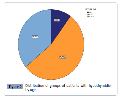
Figure 2 Indicates the distribution of groups of patients with hypothyroidism by age: 5 (10%) were 18-20 years old while 27 (54%) were aged 31-50 and: 18 (36%) were 51-78 year old.


Health-Science-journal-Distribution
 

Figure 2:Distribution of groups of patients with hypothyroidism by age.

Discussion

During this research we have been more focusing to the link between serum of Vitamin-D levels and thyroid function tests in patients with eutiroids and hypothyroids. In this study there were a total of one hundred (100) subjects. Fifty (50) of these had hypothyroidism. From (50) hypothyroid subjects: 16 (32%) were male, while 34 (68%) were females. This value indicates that the prevalence of the disease is greater in women than in men (graph 1). This matches [7]. Their study stated that hypothyroidism is a widespread disease of specific organs and affects 2 - 5% of the population with significant variability between sex (ie, female 5-15% and Men 1-5%).The same finding was confirmed by [8], who reported that the prevalence of hypothyroids is common in women. This may be due to the fact that women are more susceptible to autoimmune diseases than men. As it had been recently reported, many autoimmune disorders tend to affect women during long periods of stress, such as pregnancy, or during a hormonal change.

Also from (50) hypothyroid entities: 5 (10%) were 18-20 years old while 27 (54%) were 31-50 years old and: 18 (36%) were 51-78 years old. Recent results and data from observation studies can be said that serum TSH levels increase in older people. However, very mild TSH rising in older individuals may not reflect subclinical thyroid dysfunction, but be more than a normal consequence of aging [9].

An analysis from the Longitudinal Baltimore Aging study has found that changes in thyroid function tests are common, especially in older age groups, and medium regression is partially responsible for this finding. Importantly, changes in TSH and FT4 over a 7-year period were accompanied by increased mortality [10]. The results of the thyroid function indicate that the TSH in the group of patients with hypothyroidism has increased significantly, especially in the TPO-antibodies-positive than in the negative TPO antibodies compared to the control group. FT4 has been significantly reduced in TPO positive antibodies than in negative TPO antibodies compared to the control group. This matches the study done by [11], which found that autoimmunity is closely linked to thyroid function and an increase in autoimmunity is directly linked to the deterioration of thyroid function as they are seen increasing TSH levels in Positive anti-TPO patients.

In contrast, vitamin 25-oh D levels in the group of patients with hypothyroidism, in the positive TPO antibodies was (10.38 ± 4.45ng/ml) and in the group of negative TPO antibodies was (18.79 ± 8.93ng/ml) while compared with the control group (64.92 ± 161.08ng/ml) were significantly lower. This is compatible with the study done by (Idiculla J et al, 2018) that the level of vitamin 25-Oh D in patients with hypothyroidism was significantly lower than in eutroid controls and that anti-TPO positive patients had lower levels of Vitamin-D25- Oh compared to the negative group to anti-Tpo.

This study compares subjects that have Vitamin-D 25-Oh and those with insufficiency, thus showing an increase in TSH and anti-tpo among those with Vitamin-D 25-Oh. In contrast, the FT4 was lower in the group with Vitamin-D deficiency 25-OH than in the group with Vitamin-D 25-Oh. This relationship is confirmed by the findings of (Richards B, 2008) in its experimental study that explored the effect of Vitamin-D deficiency on the thyroid gland. In this study, he reported that Vitamin-D deficiency contributed to the possibility of lowering thyroid hormones.

The study done by [12-18], suggests that Vitamin-D deficiency is more linked to the antitroid antibody titrine than thyroid function itself in humans.

Conclusion and Recommendations

From the research results we can conclude as follows: Vitamin-D levels 25-OH were significantly reduced in patients with hypothyroidism. Patients with anti-TPO positive suffered more from hypovitaminosis D than those with negative anti- TPOs. Vitamin-D deficiency was associated with the presence of autoantibody of the thyroid and abnormal thyroid functions.

Considering the importance of Vitamin-D, we recommend: Further research fully illuminate the role of Vitamin-D levels in autoimmune thyroiditis, and provide knowledge of Vitamin-D efficiency and safety as a therapeutic tool for Aitd.

Acknowledgement

We would like to acknowledge the contribution of proofreading the article of Prof. Ass.Dr. Mulaim Kacka from the Heimerer College, for the critical review of this manuscript.

Statement of Interests

The authors declare no conflict of interest.

References

  1. Stathatos N, Daniels GH (2012) autoimmune thyroid disease. Curr Opin Rheumatol 24:70–5.
  2. Indexed at, Google Scholar, Crossref

  3. Wang J, Lv S, Chen G, Gao C, He J, et al. (2015) Meta-analysis of the association between vitamin D and autoimmune thyroid disease. Nutrients 7:2485–2498.
  4. Indexed at, Google Scholar, Crossref

  5. Chaker L, Bianco AC, Jonklaas J, Peeters RP (2017) hypothyroidism. Lancet 390:1550–1562.
  6. Indexed at, Google Scholar, Crossref

  7. AG Madariaga, Santos PS, Guillén GF, Galofré JC (2014) the incidence and prevalence of thyroid dysfunction in Europe a meta-analysis. J Clin Endocrinol Metab 99:923-931.
  8. Indexed at, Google Scholar, Crossref

  9. Vanderpump MP (2011) the epidemiology of thyroid disease.Br Med Bull 99:39-51.
  10. Indexed at, Google Scholar, Crossref

  11. Almandoz JP, Gharib H (2012) Hypothyroidism etiology diagnosis and management. Med Clin North Am 96:203-221.
  12. Indexed at, Google Scholar, Crossref

  13. Amaya AJ, Rojas-Villarraga A, Anaya JM (2014) cardiovascular disease in the antiphospholipid syndrome. Lupus 23:1288-91.
  14. Google Scholar, Crossref

  15. Meng, Zhaowei (2015) Gender and age impacts on the association between thyroid function and metabolic syndrome in Chinese. Medicine 94: e2193.
  16. Indexed at, Google Scholar, Crossref

  17. Aggarwal N, Razvi S (2013) Thyroid and aging or the aging thyroid? An evidence-based analysis of the literature. J Thyroid Res 2013:481287.
  18. Indexed at, Google Scholar, Crossref

  19. Mammen JS, McGready J, Ladenson PW, Simonsick EM (2017) Unstable thyroid function in older adults is caused by alterations in both thyroid and pituitary physiology and is associated with increased mortality. Thyroid 27:1370–1377.
  20. Indexed at, Google Scholar, Crossref

  21. Calcaterra V, Montalbano C, Miceli E, Luinetti O, Albertini R, et al. (2020) Antigastric parietal cell antibodies for autoimmune gastritis screening in juvenile autoimmune thyroid disease. J Endocrinol Invest 43:81-86.
  22. Indexed at, Google Scholar, Crossref

  23. Tamer G, Arik S, Tamer I, Coksert D (2011) Relative vitamin D insufficiency in Hashimoto's thyroiditis. Thyroid 21:891-896.
  24. Indexed at, Google Scholar, Crossref

  25. A Gil, Plaza-Diaz J, Mesa MD (2018) Vitamin D Classic and Novel Actions. Ann Nutr Metab 72:87-95.
  26. Indexed at, Google Scholar, Crossref

  27. Doohe Kim (2017) The Role of Vitamin D in Thyroid Diseases. Int J Mol Sci 18:1949.
  28. Indexed at, Google Scholar, Crossref

  29. General Information/Press Room [Internet]. American Thyroid Association.
  30. Idiculla J, Prabhu P, Pradeep R, Khadilkar K, Kannan S (2018) Vitamin D and primary hypothyroidism: Is there an association? Thyroid Res Pract 15:34.
  31. Google Scholar

  32. National Health Service (NHS). Vitamin D. 2022.
  33. Richards B (2008) Low vitamin D contributes to thyroid problems. Health news.
  34. Google Scholar

Citation: Morina M, Bilalli A, Salihu E, Kastrati K, Krasniqi M, et al. (2024) Relationships between Vitamin-D Levels in Serum and Tests of Thyroid Function in Eutroid and Hypothyroid in Patients with Anti -TPO Elevated in the Municipality of Prishtina. Health Sci J. Vol. 18 No. 5: 1135.