Theresa NH Leung1* and Na Xie2
1Department of Pediatrics and Adolescent Medicine, Faculty of Medicine, University of Hong Kong, Hong Kong SAR, People's Republic of China
2Department of Medical Imaging, University of Hong Kong-Shenzhen Hospital, Shenzhen, People's Republic of China
Corresponding Author:
Theresa NH Leung
Department of Paediatrics and Adolescent Medicine, Faculty of Medicine, University of Hong Kong, Hong Kong SAR, People's Republic of China
Tel: +852 66492317
E-mail: leungnht@hku.hk
Received date: 02 June 2016; Accepted date: 03 June 2016; Published date: 05 June 2016
Citation: Leung TN, Xie N. Scimitar Syndrome with Dilated Azygos Vein and Interrupted Inferior Vena Cava (IVC). Ann Clin Lab Res. 2016, 4:2.
A 3 year-old girl with recurrent chest infections had chest radiograph showing the typical “Turkish sword” sign which could be found in 70% in the childhood-adult group of Scimitar syndrome, a rare form of partial anormalous pulmonary venous drainage (PAPVD) associated with hypoplastic lung. The anomalous pulmonary veins drain into the systemic venous system such as inferior vena cava (IVC), right atrium, hepatic or portal vein.
Keywords
Diagnostic imaging, Scimitar syndrome, Azygos vein, Pediatrics
Image Case
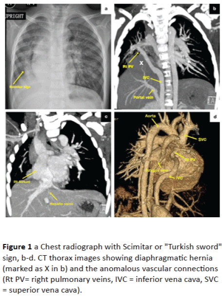
A 3 year-old girl with recurrent chest infections had chest radiograph (Figure 1a) showing the typical “Turkish sword” sign which could be found in 70% in the childhood-adult group of Scimitar syndrome, a rare form of partial anormalous pulmonary venous drainage (PAPVD) associated with hypoplastic lung [1]. The anomalous pulmonary veins drain into the systemic venous system such as inferior vena cava, right atrium, hepatic or portal vein [1].

Figure 1: a Chest radiograph with Scimitar or "Turkish sword" sign, b-d. CT thorax images showing diaphragmatic hernia (marked as X in b) and the anomalous vascular connections (Rt PV= right pulmonary veins, IVC = inferior vena cava, SVC = superior vena cava).
The reconstructed Computerized Tomography (CT) images of thorax in this patient demonstrated a few unusual features of the rare condition:
1) Anomalous right pulmonary veins joined the inferior vena cava (IVC) was interrupted after coursing through the liver (Figure 1b).
2) Azyos and hemiazygos veins were dilated (Figure 1d).
3) The hepatic vein drained directly to right atrium (Figure 1c).
Similar to two reported cases [2,3], the IVC was interrupted and the dilated azygos and hemiazygos veins received venous blood from the anomalous scimitar vein and all infradiaphragmatic systemic return except the hepatic vein which was draining to the right atrium separately. Scimitar syndrome is also classified as “combined pulmonary and vascular lesions” in the group of congenital pulmonary malformations [4]. This patient had hypoplasia of the right lung, right middle lobe and lower lobe bronchi, and a small diaphragmatic hernia involving part of the liver (marked with X in Figure 1b). Scimitar syndrome with various complex vascular connections and pulmonary malformations are described in literature, however, the embryological development of the congenital abnormalities is still not well defined.
Ethical Approval
Formal consent is not required for retrospective review and no identifiable detail of the individual is included in the manuscript and figures.
9492
References
- Cicek S (2014) Scimitar syndrome: the Turkey Sword sabre. Semin Thorac Cardiovasc Surg Pediatr Card Surg Ann 17:56-61.
- Greenberg SB (2008) Combined Scimitar Syndrome and Interruption of the Inferior Vena Cava Causing Mega-azygous and Hemiazygous Veins. Pediatr Cardiol 29:243–244.
- Celik M (2012) Scimitar Syndrome and Azygos Continuation of the Inferior Vena Cava Diagnosed in an Adult-An Unusual Association.Congenit Heart Dis7: 85-88.
- Lee E, Dorkin H, Vargas SO (2011) Congenital Pulmonary Malformations in Pediatric Patients: Review and Update on Etiology, Classification, and Imaging Findings. Radiol Clin N Am49 :921–948.


