Introduction
|
| As defined by Stewart [1], spontaneous regression of cancer consists in the complete disappearance or partial shrinkage of a malignant neoplasm without appropriate treatment. Apparently more common for renal cell carcinoma, non-Hodgkin lymphoma and leukaemia, neuroblastoma and melanoma, spontaneous regression of neoplasms has been reported for colorectal cancer only in 21 cases from 1900 up to 2005, as published in an extensive review by Abdelrazeq [2]. Of these, only nine cases account for regression of liver metastasis. After publication of Abdelrazeq’s review, some cases of spontaneous regression of colorectal cancer have been reported, but it is still clear that this remains a particularly rare event [3-5]. In spite of being a rather controversial subject among the scientific community, spontaneous regression of malignant tumours is undoubtfully a fascinating phenomenon that has intrigued many clinicians and has led a number of researchers to believe that by understanding the physiological mechanisms behind it, we might develop new key targets for the treatment and prevention of cancer [6]. |
Methods
|
| A 51-year-old-woman, submitted to a left colectomy, splenectomy and total hysterectomy (R0) followed by adjuvant chemotherapy (12 FOLFIRI cycles) for treatment of a moderately differentiated adenocarcinoma of the splenic angle of the colon, staged pT3,N1,M0 (stage III of the AJCC/UICC). Six years later, the patient was referred to our unit with two unspecific hepatic lesions, probably colorectal liver metastases. These lesions were not present in none of the precedent imaging tests (at least one scan a year) undertaken after colectomy. She reported a medical history of hypertension, type 2 diabetes and depressive syndrome; she was asymptomatic, with no relevant findings in the objective examination. |
Results
|
| Blood tests revealed a slight elevation in ALT (40 U/L; N: <34) and GGT (49 U/L; N: 48), with values for AST and AF within the normal range. |
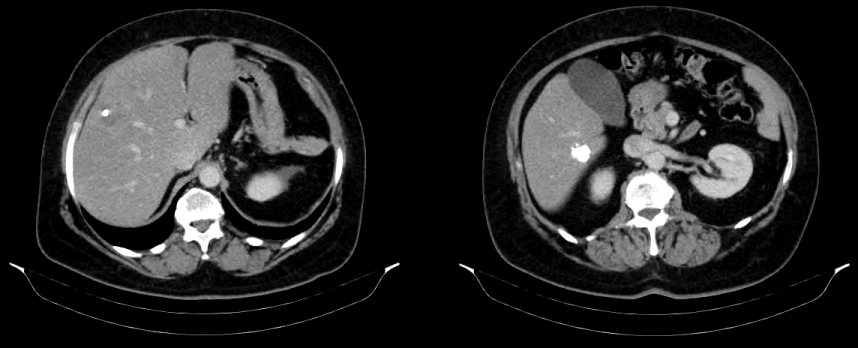
| The 6-year post-operative abdominal CT scan (Figure 1) showed an hepatic lesion in the right lobe (segment VIII), lobulated, with oval shape, denser than the normal hepatic parenchyma in precontrast phase, hypovascular after IV contrast injection, grossly calcified, measuring 57 mm in its largest axis; it also showed another grossly calcified nodular lesion in segment V/VI. |
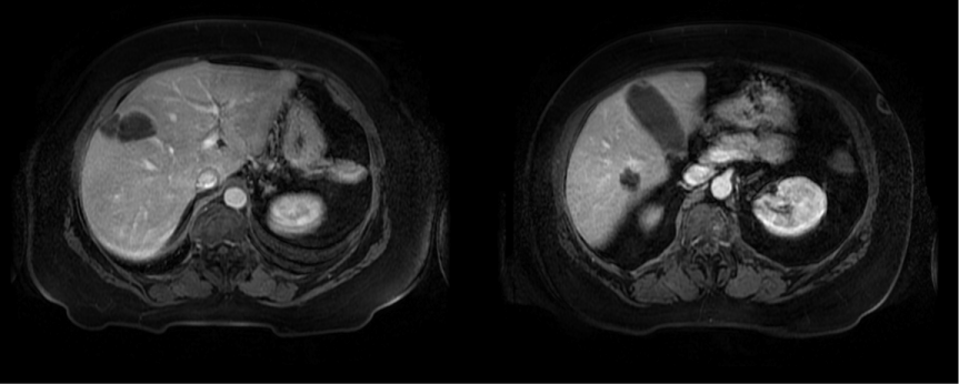
| For better assessment of these lesions, an abdominal MRI scan was requested (Figure 2), which showed a nodular formation in the segment VIII of the liver, with 5.5 × 2.8 cm in size, slightly hypointense in T1 and isointense in T2, with peripheral ring-like contrast-enhancement after administration of IV gadolinium, with small punctiform images suggestive of calcification process, conditioning minor retraction of the Glisson’s capsule; it also showed another vaguely nodular lesion, with 2 × 1.6 cm in size, in segments V/VI, with similar signal and contrast-enhancement characteristics to those of the first lesion. At this time, the tumour markers related to colorectal cancer (CEA and CA 19-9) were within normal range. |
| In view of the patient’s medical history and taking into account that there were two new lesions with typical imagiological characteristics, the diagnosis of liver metastases from colorectal cancer became highly suggestive. Solitary necrotic nodules of the liver are an exceedingly rare entity, mostly presented as unique nodules and are never bigger than 5 cm in diameter [7]. The lesions detected in the CT and MRI scans did not appear to have a cystic nature, therefore excluding the diagnosis of mucinous tumours, which could well become necrotic. |
| Considering the excellent performance status of the patient, a right hepatectomy was performed after confirmation of the preoperative imagiological findings by a per-operative ultrasound scan. |
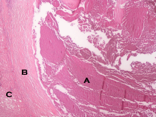
| No complications occurred in the postoperative period, and the patient was discharged on the 7th post-operative day. The pathological examination (Figure 3) revealed two nodules, completely necrotic, with no viable tumour. |
| The patient is presently on the ninth post-operative month, and remains asymptomatic with no clinical, biological or imagiological signs of relapse. |
Discussion
|
| Everson [8] considers that the concept of spontaneous tumour regression may encompass regression of a primary tumour or a metastatic tumour with or without histological confirmation of its malignancy; or the regression of presumptive metastases diagnosed through radiological methods. These later types of regression, in which there is no histological proof of malignancy, can report to rather questionable cases, providing divergent opinions amongst researchers, regarding the criteria that should be used to accurately identify spontaneous remission of cancerous lesions. This fact explains why its true incidence remains unclear, even though it has been suggested that it can be estimated between 1:80.000 and 1:100.000 cancer cases [9]. Furthermore, cases of regression seem to be particularly scarce in cases of colorectal cancer when compared to other types of cancer, particularly when we consider that colorectal cancer represents 15% of all malignancies but accounts for a mere 2% of all the reported cases of spontaneous cancerous remissions [2]. Abdelrazeq [2] suggests that cancer types with lower incidence may be less common precisely because they tend to spontaneously regress more often. Another possible explanation for this discrepancy is that more common tumours, such as bowel cancer, are also more promptly tackled [3]. |
| A recent review of cases of spontaneous regression of colorectal cancer has concluded that it typically occurs in males, more often for rectal than for colonic cancer, with a predominance in the distal relatively to the proximal colon (ratio of 3:1); and that it is exceedingly rare for extra-peritoneal metastases to spontaneously regress [2]. Moreover, liver metastases have been more often reported to regress in cases of metastatic renal cell carcinoma than in other kinds of metastatic liver malignant lesions [10]. |
| A commonly suggested etiological factor for metastatic regression is the removal of the primary tumour. It has been proposed that the surgical excision of the primary lesion may disseminate cancerous cells through both lymphatic and vascular vessels which, in turn, are responsible for a sudden increase of tumour antigens in the blood stream; high enough to stimulate an innate anti-cancerous immune response [2]. This, however, seems to be an unlikely explanation for the involution of liver metastases in the case reported in this article, as the surgical removal of the primary tumour occurred six years before the detection of the metastatic lesions, which means that they shouldn’t be present at the time of that tumour antigen peak, triggered by the surgical procedure. |
| In this case, the etiology of the spontaneous regression remains unclear. Another mechanism proposed as the basis for tumour regression is spontaneous necrosis, either by inhibition of angiogenesis or by rapid growth of the tumour. Other causes seem more unlikely. Abdelrazeq emphasizes the role of severe sepsis with prolonged high fever as a non-specific factor that stimulates the immune response in a way that could contribute to cancerous remission [2]. Yet, there was no such septic event observed in our patient. A hormonal-related response also seems improbable, as colorectal cancer is less prone to be influenced by endocrine changes as are breast and prostatic cancer, for instance [11]. Genetic and epigenetic mechanisms have also been suggested as possible factors. Another yet more controversial basis for tumour remission are psychological factors, as demonstrated by patients whose tumour underwent spontaneous regression after intensive psychotherapy [2,9]. |
| It is likely that the etiology of spontaneous tumour regression is multifactorial. But due to the rarity of the event, it is quite difficult to reach a statistically significant number of cases that may allow us to assert its true cause. |
Conclusion
|
| Spontaneous tumour regression is an exceedingly rare event. However it may provide a decisive insight on critical physiological processes that could be used in the future to treat or prevent different kinds of cancer. |
Figures at a glance
|
 |
 |
 |
| Figure 1 |
Figure 2 |
Figure 3 |
|
| |
| |


