Kiri?o?lu M Ü1*, Ösün A2, Atay B1 and Samanc?o?lu A1
Buca Seyfi Demirsoy State Hospital, Department of Neurosurgery ?zmir, Turkey
Dumlupinar University Medical School, Department of Neurosurgery, Kütahya, Turkey
Corresponding Author:
Kiri?o?lu Mehmet Ünal MD
Buca Seyfi Demirsoy State Hospital
Department of Neurosurgery ?zmir, Özmen Cad. No:145
Buca/?zmir, Turkey
E-mail: mukirisoglu@yahoo.com
Citation: Kiri?o?lu M Ü, Ösün A, Atay B, Samanc?o?lu A. Supratentorial Intraparenchymal Hemorrhage after Spinal Meningioma Surgery. J Biomedical Sci. 2015, 4:2. doi:10.4172/2254-609X.10009
Copyright: © 2015 Kiri?o?lu M Ü. This is an open-access article distributed under the terms of the Creative Commons Attribution License, which permits unrestricted use, distribution, and reproduction in any medium, provided the original author and source are credited.
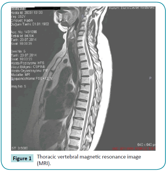
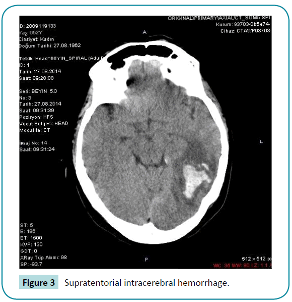
A 52-year old woman was admitted with radiating left leg pain and weakness while walking for approximately six months. Neurological examination revealed left lower monoparesis (3/5) and T8 hypoesthesia with bilateral Babinski sign. Thoracic vertebral magnetic resonance image (MRI) demonstrated a mass lesion causing a nearly total occlusion of the spinal canal (Figure 1). We removed the tumor totally in prone position via T5 and 6 laminectomy. The neuropathological examination showed it to be meningotheliamatous meningioma. Cerebrospinal fluid (CSF) leakage during the operation continued postoperatively through the subfascial drain though the dura was closed properly. At the second day of the operation, the patient began to complain about headache. Though we didn’t observe additional neurological deficit, CT scan demonstrated a right parietooccipital intracerebral hemorrhage (Figure 2). The patient was treated conservatively. We stopped subfascial drainage and her complaints regressed without any additional neurological defisit [1] (Figure 3).

Figure 1: Thoracic vertebral magnetic resonance image (MRI).

Figure 2: CT scan demonstrated a right parietooccipital intracerebral hemorrhage.

Figure 3: Supratentorial intracerebral hemorrhage.
Supratentorial intracerebral hemorrhage in post spinal intradural surgery patients are extremely rare. The mechanism of intracranial hemorrhage is commonly explained by intracranial hypotension and traction of cerebral veins due to CSF loss. A similar mechanism also explains the headache due to lumbar puncture (LP) and spinal anesthesia. Apart from the patients developing neurological deficits, most cases benefit from conservative therapy. Avoiding CSF loss, plasma volume expansion and applying epidural patch for persistant headache are treatment choices. The patients headache in our case improved in a few days after the removal of subfascial drain [2-4].
In order to determine the real frequency of intracerebral hemorrhage due to CSF loss resulting from spinal surgery, spinal anesthesia, and LP we recommend brain imaging studies to be done in cases with headache even without neurological deficits as in our case.
7214
References
- Gilberto Ka Kit Leung, Johnny Ping Hon Chan (2014) Supratentorial intraparenchimal haemorrhages during spine surgery: case report. J Korean Neurosurg soc 55:103-105.
- Çak?r CÖ, Balo?lu M, Ataizi ZS (2014) Supratentorial hemorrhage following spinal tumor surgery: Konuralp T?p Dergisi 6: 53-56.
- Moradi M, Shami S, Farhadifar F, Nesseri K (2012) Cerebral subdural hematoma following spinal anesthesia: Report of two cases: Hindawi journals2012: 4.
- Greenberg MS(2010) Headache in:Greenberg M S (ed), Handbook of Neurosurgery, Thieme, 2010:57-59


