Hisashi Ito1*, Jordi Rumià2, Shigeru Fukutake1, Kazuaki Yamamoto3, Toshio Yamaguchi4, Takaomi Taira5 and Tetsumasa Kamei1
1Department of Neurology, Shonan Fujisawa Tokushukai Hospital, Fujisawa, Japan
2Department of Functional Neurosurgery, ResoFUS ALOMAR Barcelona, University of Barcelona, Barcelona, Spain
3Department of Neurosurgery, Shonan Kamakura General Hospital, Kamakura, Japan
4Research Institute of Diagnostic Imaging, Shin-Yurigaoka General Hospital, Kawasaki, Japan
5Department of Neurosurgery, Tokyo Women’s Medical University, Tokyo, Japan
*Corresponding Author:
Dr. Hisashi Ito
Department of Neurology
Shonan Fujisawa Tokushukai Hospital
251-0041, Fujisawa, Japan
Tel: +81-466-35-1177
Fax: +81-466-35-1300
E-mail: hisashi.ito@tokushukai.jp
Received Date: February 26, 2019; Accepted Date: March 15, 2019; Published Date: March 20, 2019
Citation: Ito H, Rumià J, Fukutake S, Yamamoto K, Yamaguchi T, et al. (2019) The Efficacy of Bisphosphonate to Increase the Skull Density Ratio of MRI-guided Focused Ultrasound Candidates with Brain Disorders. J Neurol Neurosci Vol.10 No.2:293. DOI: 10.36648/2171-6625.10.2.293
Background: Skull density ratio (SDR) is one of the key factors for the success of MRI-guided focused ultrasound (MRgFUS) treatment for brain diseases. We examined the efficacy of alendronate (Aln) to improve a SDR value.
Materials and Methods: The subjects were 6 Parkinson’s disease patients (2 men, 4 women, 70.7 ± 7.7 years old) and 1 essential tremor patient (1 man, 81 years old). Despite of desire to MRgFUS treatment, we could not schedule their treatments because their SDR values were too low. As they coexisted with untreated osteoporosis, we administered 35 mg of Aln weekly and followed their SDR values every 3 months.
Results: The SDR value elevated in 4 patients following the administration of Aln and we could perform MRgFUS treatment successfully. No adverse reactions related with Aln were observed.
Conclusion: Although it will take several months, Aln may be a useful option for MRgFUS candidates with a low SDR value.
Keywords
Skull density ratio; Brain diseases; Parkinson's disease
Introduction
Transcranial MRI–guided focused ultrasound (MRgFUS) is a novel neurosurgical tool based on the technology by Fry brothers [1]. It consists of high intensity ultrasound for ablation, MR imaging for confirming target location, and MR thermography for real-time monitoring of thermal process. Compared to previous neurosurgical interventions, MRgFUS is characterized with minimum invasiveness as it requires neither burr hole opening nor device implantation. MRgFUS has been applied for several neurological or psychological disorders, including Parkinson’s disease (PD) and essential tremor (ET) [2-8]. There are two key factors determining the success of MRgFUS treatment for brain diseases: the skull volume and skull density ratio (SDR) [9]. Here we report a preliminary research on the efficacy of alendronate (Aln), one of the popular bisphosphonates for osteoporosis, to elevate the SDR value in patients of PD and ET.
Materials and Methods
This is a preliminary, open labeled and retrospective study whose objects were 6 PD patients (2 men, 4 women, 70.7 ± 7.7 years old) and 1 ET patient (1 man, 81 years old). Demography of patients was shown in the Table 1. All the patients desired to perform MRgFUS treatment, which had been approved by the Review Board of Tokushukai Medical Alliance or University of Barcelona. They provided a written informed consent in accordance with the Declaration of Helsinki before treatment commenced. However, we had to suspend their treatments because of low SDR values (exclusion criteria: ≤ 0.30 in Japan, ≤ 0.40 in EU). As they had untreated osteoporosis diagnosed with a standard examination, we administered Aln 35 mg weekly and examined their SDR values every 3 months.
| No. |
Age |
Gender |
SDR at baseline |
Site |
Disease |
Target |
| 1 |
56 |
M |
0.35 |
UB |
PD |
Vim |
| 2 |
78 |
F |
0.26 |
SFTH |
PD |
GPi |
| 3 |
80 |
M |
0.25 |
SFTH |
ET |
Vim |
| 4 |
70 |
F |
0.24 |
SFTH |
PD |
GPi |
| 5 |
72 |
F |
0.29 |
SFTH |
PD |
Vim |
| 6 |
75 |
F |
0.29 |
SFTH |
PD |
Vim |
| 7 |
73 |
M |
0.27 |
SFTH |
PD |
Vim |
UB: University of Barcelona; SFTH: Shonan Fujisawa Tokushukai Hospital
Table 1: Demography of patients.
Results
We followed the patients for 3 to 9 months of the administration of Aln. The SDR value is elevated gradually in 4 patients, though it reached a plateau in 2 patients (Figure 1). For these patients, we performed MRgFUS Vim thalamotomy or GPi pallidotomy and archived good outcomes [7]. We did not observe Aln-related adverse reactions at all.
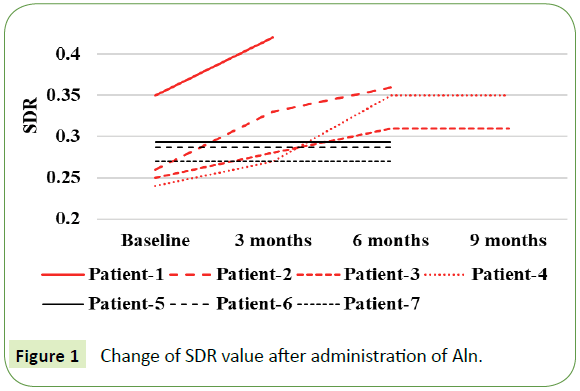
Figure 1: Change of SDR value after administration of Aln.
Discussion
Transcranial MRgFUS is an innovative technology for several psychological and neurological disorders including PD and ET. The main advantages of MRgFUS are minimum invasiveness and the immediate onset of therapeutic effect. Most of the adverse events are mild and transient [2,5-8]. The primary therapeutic targets for PD and ET are ventral intermediate nucleus (Vim) for tremor [3,4,6] and globus pallidus internus (GPi) for motor-fluctuations [7,8]. In addition, the effectiveness and safety of pallidothalamic tractotomy for tremor and dyskinesia had been reported [5].
The skull is a marked barrier to ultrasonic energy transmission. SDR, the mean value for the ratio of Hounsfield units of marrow and cortical bone, which reflects the amount of ultrasonic energy that can penetrate the skull effectively, is one of the key factors needed to be taken into consideration for a successful MRgFUS procedure for brain disorders, and it shows a positive correlation with maximal temperature in the target lesion [9]. Therefore, optimizing a SDR value may be useful for MRgFUS candidates with a low SDR value, and it might be helpful to expand the patients indicated for MRgFUS.
Moreover, ultrasound elements tend to have a high incident angle with the skull and the number of elements which deliver energy effectively to the target is limited in MRgFUS GPi pallidotomy compared with Vim thalamotomy or pallidothalamic tractotomy because the GPi target locates laterally. Thus, high SDR value is critical especially in MRgFUS GPi pallidotomy. However, the appropriate method to elevate a SDR value has yet to be elucidated.
Aln is one of the third-generation bisphosphonates. Although the association between SDR and osteoporosis is not clear, Aln was previously reported to increase the degree and uniformity of bone matrix mineralization and decrease the porosity of cortical bone [10]. Aln could be a useful option for MRgFUS candidates with a low SDR value; however, the presence of non-responders and two patients reaching a plateau indicates the limitation of the efficacy of Aln and we have to consider that it takes several months for achieving enough value of SDR. In addition, bisphosphonates other than Aln, RANKL inhibitor, and recombinant human parathyroid hormone might have some potential to impact positively on a SDR value. Further investigations concerning the efficacy of Aln and other drugs for optimizing a SDR value are necessary.
Conclusion
Aln could elevate a SDR value in some MRgFUS candidates with brain disorders.
Acknowledgement
We would like to thank Mr. Shinya Tanuma (Daiichi-Sankyo) for his advices to pathomechanism and a current treatment of osteoporosis.
Sources of Financial Support
No funding received for this work.
Conflict of Interest
There are no conflicts.
24238
References
- Fry WJ, Mosberg WH, Barnard JW, Fry FJ (1954) Production of focal destructive lesions in the central nervous system with ultrasound. J Neurosurg 11: 471-478.
- Weintraub D, Elias WJ (2017) The emerging role of transcranial Magnetic Resonance Imaging–guided focused ultrasound in functional neurosurgery. Mov Disord 32: 20-27.
- Lipsman N, Schwartz ML, Huang Y, Lee L, Sankar T, et al. (2013) MR-guided focused ultrasound thalamotomy for essential tremor: a proof-of-concept study. Lancet Neurol 12: 462-468.
- Elias WJ, Lipsman N, Ondo WG, Ghanouni P, Kim YG, et al. (2016) A randomized trial of focused ultrasound thalamotomy for essential tremor. N Engl J Med 375: 730-739.
- Magara A, Bühler R, Moser D, Kowalski M, Pourtehrani P, et al. (2014) First experience with MR-guided focused ultrasound in the treatment of Parkinson's disease. J Ther Ultrasound 2: 11.
- Schlesinger I, Eran A, Sinai A, Erikh I, Nassar M, et al. (2015) MRI guided focused ultrasound thalamotomy for moderate-to-severe tremor in Parkinson’s disease. Parkinson’s Dis 2015: 219149.
- Na YC, Chang WS, Jung HH, Kweon EJ, Chang JW (2015) Unilateral magnetic resonance-guided focused ultrasound pallidotomy for Parkinson disease. Neurology 85: 549-551.
- Ito H, Taira T, Fukutake S, Yamamoto K, Baba Y, et al. (2018) Magnetic resonance imaging-guided focused ultrasound unilateral pallidotomy for Parkinson’s disease: A case report. Int J Case Rep 2: 07.
- Chang WS, Jung HH, Zadicario E, Rachmilevitch I, Tlusty T, et al. (2016) Factors associated with successful magnetic resonance-guided focused ultrasound treatment: efficiency of acoustic energy delivery through the skull. J Neurosurg 124: 411-416.
- Roschger P, Rinnerthaler S, Yates J, Rodan GA, Fratzl P, et al. (2001) Alendronate increases degree and uniformity of mineralization in cancellous bone and decreases the porosity in cortical bone of osteoporotic women. Bone 2: 185-191.

