Yassine Echchikhi*, Asmae Touil, Sara Elabbassi, Tayeb Elkebdani and Noureddine Benjaafar
Department of Radiation Oncology, National Institute of Oncology, Mohammed V University, Rabat, Morocco
- *Corresponding Author:
- Yassine Echchikhi
Department of Radiation Oncology
National institute of Oncology
Mohamed V University, Rabat, Morocco
Tel: +212662786099
E-mail: eyassine12@hotmail.com
Received Date: July 22, 2017; Accepted Date: August 09, 2017; Published Date: August 28, 2017
Citation: Echchikhi Y, Touil A, Elabbassi S, Elkebdani T, Benjaafar N (2017) Undifferentiated Lymphoepithelial Carcinoma of Cervix: A Rare Case Report. Arch Can Res Vol.5:No.3:150. DOI: 10.21767/2254-6081.1000150
Keywords
Lymphoepithelial carcinoma; Cervix; Management tumors, Post-menopausal bleeding
Introduction
Lymphoepithelial carcinoma (LEC) is not common malignant tumor which composed of undifferentiated malignant epithelial cells associated characteristic lymphoid stroma. This frequently occurred in nasopharynx. Meanwhile, it can also occur in other organs like the salivary glands, particularly the parotid and submandibular glands. It is significantly associated with Epstein-Barr virus (EBV). These tumors mainly affect females in the fifth decade of life.
Case Presentation
A 50-year-old woman presented in our department with 24 months history of post-menopausal bleeding of low abundance and pelvic pain, which was increasing in intensity over 3 months. However, she denied any weight loss, fever or altered urinary or bowel habits.
On physical assessment, she was with good performance status without pallor. Vaginal examination showed an ulcer and budding tumor destroying the cervix and the upper third of vagina.
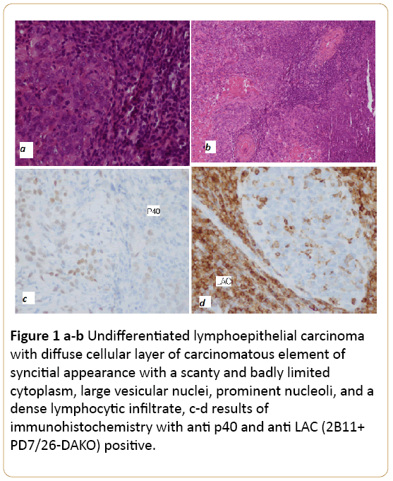
A cervical biopsy was performed the histological results with immunohistochemistry were in favor of undifferentiated lymphoepithelial carcinoma with the following characteristics. Evidently malignant tumor characterized by a diffuse cellular layer of carcinomatous element of syncitial appearance with a scanty and badly limited cytoplasm with large vesicular nuclei, prominent nucleoli, and a dense lymphocytic infiltrate in Figures 1a and 1b. The results of immunohistochemistry with anti p40 and anti LAC (2B11+ PD7/26-DAKO) were positive in Figures 1c and 1d.

Figure 1 a-b: Undifferentiated lymphoepithelial carcinoma with diffuse cellular layer of carcinomatous element of syncitial appearance with a scanty and badly limited cytoplasm, large vesicular nuclei, prominent nucleoli, and a dense lymphocytic infiltrate, c-d results of immunohistochemistry with anti p40 and anti LAC (2B11+ PD7/26-DAKO) positive.
Magnetic Resonance Imaging (MRI) showed a tumor of cervix measuring 30 mm × 22 mm × 20 mm, with evidence of parametriale invasion and clear interface with bladder and rectum (Figure 2). She was staging according to the International Federation of Gynecology and Obstetrics staging system 2009 as FIGO IIB. After multidisciplinary board meeting, patient was started on chemoradiation.

Figure 2: Tumor of cervix measuring 30 mm × 22 mm × 20 mm, with evidence of parametriale invasion and clear interface with bladder and rectum.
Discussion
Lymphoepithelial carcinoma (LEC) is a rare malignancy. Histologically, it is an undifferentiated carcinoma with an intermixed reactive lymphoplasmacytic infiltrate. The World Health Organization (WHO) has defined it as “a poorly differentiated squamous cell carcinoma or histologically undifferentiated carcinoma accompanied by a prominent reactive lymphoplasmacytic infiltrate, morphologically similar to nasopharyngeal carcinoma” [1]. It has been diagnosed in the oral cavity, oropharynx, nasal cavity, paranasal sinuses, salivary gland and in numerous other organs of the head and neck region, but it was extremely rare in the cervix. These tumors are considered to be a rare variant of squamous cell carcinoma (SCC) of the cervix. With an incidence ranging from 0.7% (west) to 5.5% (Asia), However being potentially radiosensitive and having fewer chances of nodal metastasis, they have a better prognosis [2].
Conclusion
There are only few case reports describing the cytology and the management of these tumors. Our case had a classical histopathology of LEC with syncytium of large anaplastic epithelial cells in a background of intense lymphoplasmacytic infiltrate [3,4].
The etiology and pathogenesis of LEC of uterine cervix is still unclear. While the role of EBV has been well established in the LEC of nasopharynx its role in that of cervix is controversial. Tseng et al. suggested EBV as a potential causative agent in 1997 who detected it in 73.3% cases of Asian women [5]. But its role in the western population seems less likely. In contrast, Chao et al. in 2009 suggested that human papilloma virus (HPV) and not EBV is involved in the carcinogenesis of LEC of the cervix and also that EBV sequences actually exist in the florid inflammatory stromal component of these tumors [6,7].
Mills et al, report in his paper one case was successfully treated with radiation therapy, with 10-year disease-free follow-up [8].
20453
References
- Tsang WY (2005) Lymphoepithelial carcinoma. Pathology and Genetics: Head and Neck Tumours 251: 18-19.
- Reich O, Pickel H, Pürstner P (1994) Exfoliative cytology of a lymphoepithelioma like carcinoma in a cervical smear. Acta cytologica 43: 285-288.
- Hamazaki M, Fujita H, Arata T, Takata S (1968) Medullary carcinoma with lymphoid infiltration of the uterine cervix-pathological picture of a case of cervix cancer with a favorable prognosis. Gan No Rinsho 14: 787-792.
- Hafiz MA, Kragel PJ, Toker C (1985) Carcinoma of the uterine cervix resembling lymphoepithelioma. ObstetGynecol 66: 829-831.
- Tseng CJ, Pao CC, Tseng LH, Chang CT, Lai CH, et al. (1997) LymphoepitheliomaÃÂÃÂÃÂâÂÂÃÂâÂÂÃÂâÃÂÃÂââ¬Ã
¡ÃÂâââ¬Ã
¡ÃÂìÃÂÃÂââ¬Ã
¡ÃÂâÂÂÃÂÃÂlike carcinoma of the uterine cervix. Cancer 80: 91-97.
- Chao A, Tsai CN, Hsueh S, Lee LY, Chen TC, et al. (2009) Does Epstein-Barr virus play a role in lymphoepithelioma-like carcinoma of the uterine cervix?Int J GynecolPathol 28: 279-285.
- Rathore R, Arora VK, Singh B (2017) LymphoepitheliomaÃÂÃÂÃÂâÂÂÃÂâÂÂÃÂâÃÂÃÂââ¬Ã
¡ÃÂâââ¬Ã
¡ÃÂìÃÂÃÂââ¬Ã
¡ÃÂâÂÂÃÂÃÂlike carcinoma of cervix. Diagnostic cytopathology 45: 239-242.
- Mills SE, Austin MB, Randall ME (1985) Lymphoepithelioma-like carcinoma of the uterine cervix: A distinctive, undifferentiated carcinoma with inflammatory stroma. Am J surglpathol 9: 883-889.


